 Back
BackComprehensive Study Notes: The Respiratory System
Study Guide - Smart Notes
 Tailored notes based on your materials, expanded with key definitions, examples, and context.
The Respiratory System
Introduction to the Respiratory System
The respiratory system is essential for gas exchange, supplying oxygen to body tissues and removing carbon dioxide. It supports aerobic metabolism, which is vital for cellular energy production. The system is divided into upper and lower components, each with specialized structures and functions.
Oxygen is obtained from the air via diffusion across lung surfaces.
Carbon dioxide is a metabolic waste product transported from tissues to the lungs for exhalation.
Organization and Structure of the Respiratory System
Major Components
The respiratory system is organized into conducting and respiratory portions:
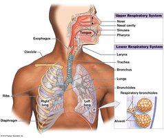
Upper respiratory system: Nose, nasal cavity, sinuses, pharynx
Lower respiratory system: Larynx, trachea, bronchi, bronchioles, alveoli
Respiratory Mucosa and Defense System
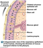
The respiratory mucosa lines the conducting portion and consists of an epithelium and underlying areolar tissue (lamina propria). It plays a critical role in filtering, humidifying, and protecting the respiratory tract from pathogens and debris.
Mucous glands in the upper tract, trachea, and bronchi secrete mucus to trap particles.
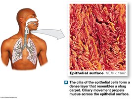
Cilia sweep mucus toward the pharynx for swallowing.
Alveolar macrophages engulf small particles that reach the alveoli.
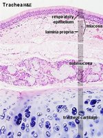
Histology of the Respiratory Tract
The respiratory tract is lined by pseudostratified ciliated columnar epithelium with goblet cells and mucous glands. The trachea also contains cartilaginous rings for support.
Lamina propria: Connective tissue layer beneath the epithelium.
Submucosa: Contains glands and blood vessels.
Tracheal cartilage: Provides structural support to keep the airway open.
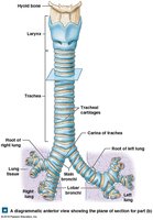
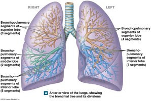
Airways and Bronchial Tree
Trachea and Bronchi
The trachea divides into right and left main bronchi, which further branch into smaller bronchi and bronchioles. The bronchial tree ensures efficient distribution of air to all regions of the lungs.
Tracheal cartilages: C-shaped rings that prevent collapse.
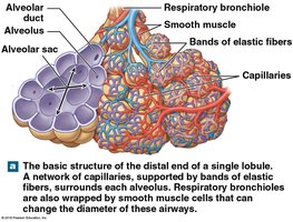
Bronchioles: Lack cartilage, dominated by smooth muscle, and regulate airflow resistance.
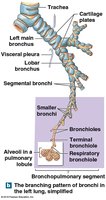
Bronchioles and Alveoli
Bronchioles branch into terminal and respiratory bronchioles, ending in alveolar sacs where gas exchange occurs. The walls of bronchioles contain smooth muscle, allowing regulation of airway diameter by the autonomic nervous system.
Bronchodilation: Sympathetic activation (β2 receptors) increases airway diameter.
Bronchoconstriction: Parasympathetic activation (M3 receptors) decreases airway diameter.
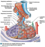
Alveolar Structure and Gas Exchange
Alveolar Epithelium
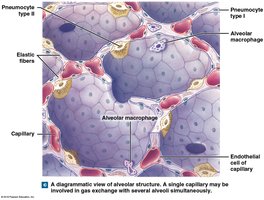
The alveoli are lined by a simple squamous epithelium composed mainly of type I pneumocytes (site of gas exchange) and type II pneumocytes (produce surfactant). Alveolar macrophages patrol the surface for debris and pathogens.
Surfactant: An oily secretion that reduces surface tension, preventing alveolar collapse.
Respiratory distress syndrome: Caused by insufficient surfactant, leading to alveolar collapse.
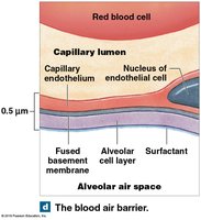
The Blood-Air Barrier
Gas exchange occurs across the blood-air barrier, which consists of three layers:
Alveolar cell layer
Fused basement membrane
Capillary endothelial layer
This barrier is extremely thin, allowing rapid diffusion of O2 and CO2.
Pleura and Lungs
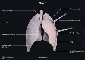
Pleural Cavities and Membranes
Each lung is surrounded by a pleural cavity lined with a double-layered serous membrane:
Parietal pleura: Lines the thoracic wall.
Visceral pleura: Covers the lung surface.
Pleural fluid: Lubricates and reduces friction between layers.
Mechanics of Breathing (Pulmonary Ventilation)
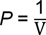
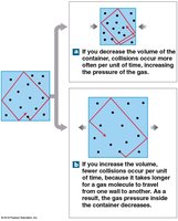
Boyle’s Law and Pressure-Volume Relationships
Boyle’s Law states that the pressure of a gas is inversely proportional to its volume, which is fundamental to understanding pulmonary ventilation:
Inspiration and Expiration
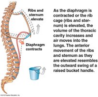
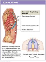
Air flows from areas of higher to lower pressure. Inspiration is always active, involving contraction of the diaphragm and external intercostal muscles. Expiration can be passive or active, depending on the involvement of accessory muscles.
Inhalation: Diaphragm contracts, thoracic volume increases, pressure decreases, air enters lungs.
Exhalation: Diaphragm relaxes, thoracic volume decreases, pressure increases, air exits lungs.
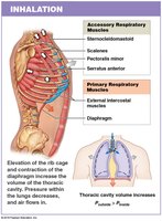
Muscles of Respiration
Primary muscles: Diaphragm (75% of air movement), external intercostals (25%).
Accessory muscles: Sternocleidomastoid, scalenes, pectoralis minor, serratus anterior (inhalation); internal intercostals, transversus thoracis, abdominal muscles (exhalation).

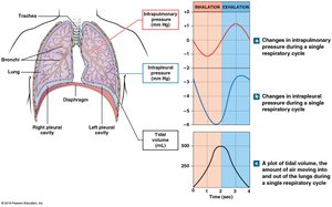
Respiratory Pressures
Intrapulmonary pressure: Pressure within alveoli; determines airflow direction.
Intrapleural pressure: Pressure in pleural cavity; always negative relative to atmospheric pressure, aiding lung expansion.
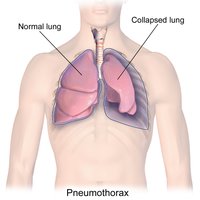
Pneumothorax and Atelectasis
Pneumothorax is the entry of air into the pleural cavity, causing lung collapse (atelectasis). It can result from trauma or alveolar rupture.
Respiratory Volumes and Capacities
Pulmonary Volumes
Tidal volume (VT): Air moved in a single breath.
Expiratory reserve volume (ERV): Air exhaled after normal exhalation.
Residual volume: Air remaining after maximal exhalation.
Inspiratory reserve volume (IRV): Air inhaled after normal inhalation.
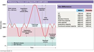
Respiratory Capacities
Inspiratory capacity: VT + IRV
Functional residual capacity (FRC): ERV + residual volume
Vital capacity: ERV + VT + IRV
Total lung capacity: Vital capacity + residual volume

Gas Exchange and Transport
Physical Principles of Gas Exchange
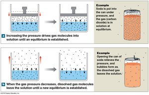
Gas exchange depends on partial pressures (Dalton’s Law) and solubility (Henry’s Law):
Dalton’s Law: Total pressure is the sum of partial pressures of individual gases.
Henry’s Law: Amount of gas dissolved in a liquid is proportional to its partial pressure.
Efficiency of Gas Exchange
Large differences in partial pressures across the blood-air barrier
Short diffusion distances
Lipid solubility of O2 and CO2
Large surface area of alveoli
Coordinated blood flow and airflow
Oxygen and Carbon Dioxide Transport
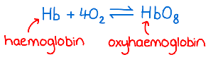
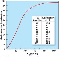
Oxygen is transported mainly bound to hemoglobin in red blood cells, forming oxyhemoglobin (HbO2):
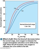
Hemoglobin saturation: Percentage of heme units with bound O2; affected by PO2, pH, temperature, and metabolic activity.
Carbon Dioxide Transport
Converted to carbonic acid (H2CO3)
Bound to hemoglobin
Dissolved in plasma
Control of Respiration
Neural Regulation
Respiratory centers in the medulla oblongata and pons regulate the rate and depth of breathing. The dorsal respiratory group (DRG) controls inspiration, while the ventral respiratory group (VRG) is active during forced breathing.
Apneustic and pneumotaxic centers: Adjust output of respiratory rhythmicity centers.
Reflex Control
Chemoreceptors: Respond to changes in PCO2, PO2, and pH.
Baroreceptors: Respond to changes in blood pressure.
Stretch receptors: Respond to changes in lung volume.
Age-Related Changes and Integration
Effects of Aging
Elastic tissue deteriorates, reducing lung compliance and vital capacity.
Arthritic changes restrict chest movement.
Emphysema is more common in older adults, especially smokers.
Integration with Other Systems
The respiratory and cardiovascular systems work together to maintain homeostatic O2 and CO2 levels in peripheral tissues.
