 Back
BackDigestive System: Structure, Function, and Processes
Study Guide - Smart Notes
 Tailored notes based on your materials, expanded with key definitions, examples, and context.
Digestive System OverviewFunctions of the Digestive System
The digestive system is responsible for processing food, breaking it down into nutrients, absorbing these nutrients, and eliminating waste. It consists of a series of organs and tissues that work together to ensure proper digestion and absorption.
Ingestion: Taking in food through the mouth.
Mechanical Breakdown: Chewing, mixing, and churning food to increase surface area for enzymes.
Digestion: Enzymatic breakdown of complex molecules into simpler forms.
Absorption: Uptake of nutrients into the bloodstream or lymphatic system.
Defecation: Elimination of indigestible substances as feces.
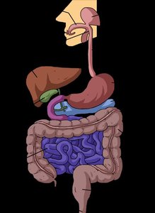
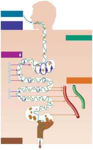
Organs of the Digestive System
The organs are divided into two groups: the alimentary canal and accessory organs.
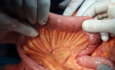
Alimentary Canal (GI Tract): Continuous tube from mouth to anus, including the mouth, pharynx, esophagus, stomach, small intestine, large intestine, and anus.
Accessory Organs: Teeth, tongue, gallbladder, salivary glands, liver, and pancreas. These organs assist in digestion by producing secretions or aiding mechanical breakdown.
Digestive Processes
The Six Essential Activities
Food processing in the digestive system involves six key steps:
Ingestion: Eating and taking food into the mouth.
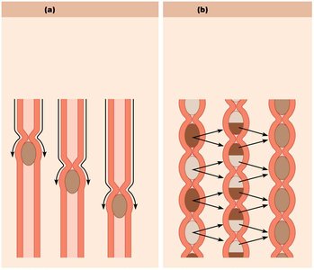
Propulsion: Moving food through the alimentary canal, including swallowing and peristalsis (alternating waves of contraction and relaxation).
Mechanical Breakdown: Physical breakdown of food by chewing, mixing with saliva, churning in the stomach, and segmentation in the intestines.
Digestion: Enzymatic breakdown of food molecules into their chemical building blocks.
Absorption: Passage of digested fragments from the GI tract lumen into blood or lymph.
Defecation: Elimination of indigestible substances via the anus.
Organization of the Digestive System
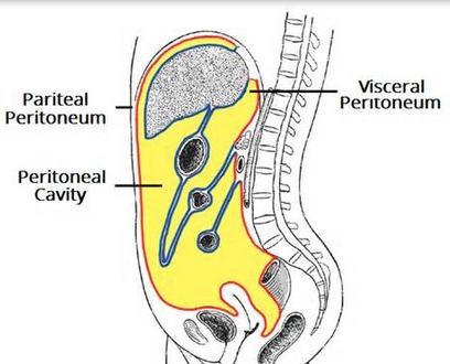
Relationship to the Peritoneum
The peritoneum is a serous membrane lining the abdominal cavity and covering the digestive organs. It consists of:
Visceral Peritoneum: Covers external surfaces of most digestive organs.
Parietal Peritoneum: Lines the body wall.
Peritoneal Cavity: Fluid-filled space between the two peritoneums, lubricating mobile organs.
Mesentery and Organ Classification
The mesentery is a double layer of peritoneum that provides routes for blood vessels, lymphatics, and nerves, holds organs in place, and stores fat. Organs are classified as:
Intraperitoneal: Located within the peritoneum (e.g., stomach, liver).
Retroperitoneal: Located outside or posterior to the peritoneum (e.g., most of pancreas, duodenum, parts of large intestine).
Histology of the Alimentary Canal
Four Basic Layers (Tunics)
All digestive organs share four basic layers:
Mucosa: Lines the lumen, secretes mucus, digestive enzymes, and hormones; absorbs end products; protects against disease.
Submucosa: Areolar connective tissue with blood and lymphatic vessels, lymphoid follicles, and nerve plexus; provides elasticity and nourishment.
Muscularis Externa: Responsible for segmentation and peristalsis; contains inner circular and outer longitudinal muscle layers; forms sphincters.
Serosa: Outermost layer, made of visceral peritoneum; replaced by adventitia in the esophagus; retroperitoneal organs have both.
Mucosa Sublayers
The mucosa consists of three sublayers:
Epithelium: Simple columnar epithelium and mucus-secreting cells in most of the tract; stratified squamous epithelium in mouth, esophagus, and anus.
Lamina Propria: Loose areolar connective tissue with capillaries for nourishment and absorption; contains lymphoid follicles (MALT).
Muscularis Mucosae: Smooth muscle producing local movements of mucosa.
Mouth and Accessory Organs
Mouth and Associated Organs
The mouth is where food is chewed and mixed with enzyme-containing saliva, beginning the process of digestion and initiating swallowing. Associated organs include:
Mouth
Tongue
Salivary glands
Teeth
Salivary Glands
Salivary glands produce saliva, which has several functions:
Cleanses the mouth
Dissolves food chemicals for taste
Moistens food and compacts it into a bolus
Begins breakdown of starch with enzyme amylase
Control of salivation: About 1500 ml/day can be produced; minor glands continuously keep the mouth moist.
Halitosis
Halitosis is bad breath caused by decomposing food particles that accumulate, allowing bacteria to flourish. The odor is mainly due to metabolic activity of anaerobic bacteria at the back of the tongue.
Summary Table: Layers of the Alimentary Canal
Layer | Main Components | Functions |
|---|---|---|
Mucosa | Epithelium, lamina propria, muscularis mucosae | Secretion, absorption, protection |
Submucosa | Areolar connective tissue, blood vessels, lymphatics, nerve plexus | Support, nourishment, elasticity |
Muscularis Externa | Inner circular and outer longitudinal muscle layers | Segmentation, peristalsis, sphincter formation |
Serosa | Connective tissue, mesothelium | Protection, structural support |
Key Terms and Concepts
Peristalsis: Propulsive movement of food through the GI tract by alternating muscle contractions.
Segmentation: Mixing movement in the intestines that breaks down food mechanically.
Mesentery: Double layer of peritoneum that supports digestive organs and provides routes for vessels and nerves.
MALT: Mucosa-associated lymphoid tissue, part of the immune defense in the GI tract.
Relevant Equations
While the digestive system is primarily anatomical and physiological, some relevant chemical equations for digestion include:
Starch breakdown by amylase:
Where starch is broken down into glucose units by the enzyme amylase.
Additional info: Academic context was added to clarify the functions and histology of the digestive system, as well as to provide a summary table and relevant chemical equation.
