 Back
BackEpithelial Tissue Types: Structure and Identification
Study Guide - Smart Notes
 Tailored notes based on your materials, expanded with key definitions, examples, and context.
Q1. Identify and describe the structure of the following epithelial tissues based on the images provided.
Background
Topic: Epithelial Tissue Classification
This question is testing your ability to recognize and describe the main types of epithelial tissues based on their cellular shape and arrangement. Understanding these tissue types is fundamental in Anatomy & Physiology, as each type has specific locations and functions in the body.
Key Terms and Concepts:
Simple epithelium: A single layer of cells.
Stratified epithelium: Multiple layers of cells.
Squamous cells: Flat, thin cells.
Cuboidal cells: Cube-shaped cells.
Columnar cells: Tall, column-like cells.
Basement membrane: A thin, fibrous layer that anchors epithelial tissue to underlying connective tissue.
Free surface: The side of the epithelial tissue exposed to the body’s exterior or to the cavity of an internal organ.
Step-by-Step Guidance
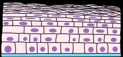
Examine each image and note the number of cell layers present. Determine if the tissue is simple (one layer) or stratified (multiple layers).
Observe the shape of the cells at the free surface (top layer): are they flat (squamous), cube-shaped (cuboidal), or tall (columnar)?
Identify the basement membrane (usually indicated by a blue line at the base of the cells) and the free surface (the exposed edge of the tissue).
Match each image to the correct epithelial tissue type based on the number of layers and cell shape:
Simple squamous epithelium
Simple cuboidal epithelium
Simple columnar epithelium
Stratified squamous epithelium
Label the cytoplasm, cell membrane, and nucleus in each diagram. Indicate the location of the basement membrane and free surface as shown in the images.



Try solving on your own before revealing the answer!
Final Answer:
Image 1: Simple squamous epithelium (single layer of flat cells)
Image 2: Simple cuboidal epithelium (single layer of cube-shaped cells)
Image 3: Simple columnar epithelium (single layer of tall, column-like cells)
Image 4: Stratified squamous epithelium (multiple layers of flat cells at the surface)
Each tissue type is identified by the number of layers and the shape of the cells at the free surface. The basement membrane is the blue line at the base, and the free surface is the exposed edge.
