 Back
BackFundamentals of the Nervous System and Nervous Tissue – Study Notes
Study Guide - Smart Notes
 Tailored notes based on your materials, expanded with key definitions, examples, and context.
Fundamentals of the Nervous System and Nervous Tissue
Overview of the Nervous System
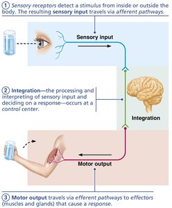
The nervous system is the master controlling and communicating system of the body. It uses electrical and chemical signals to coordinate rapid, specific, and immediate responses to internal and external stimuli.
Functions: Sensory input, integration, and motor output.
Organization: Divided into the Central Nervous System (CNS) and Peripheral Nervous System (PNS).
Divisions of the Nervous System
The nervous system is structurally and functionally divided to efficiently process and respond to information.
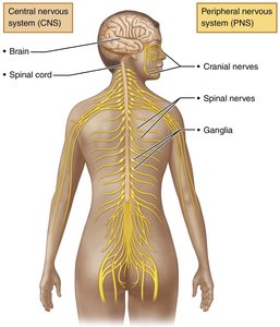
Central Nervous System (CNS): Consists of the brain and spinal cord; serves as the integration and control center.
Peripheral Nervous System (PNS): Composed of cranial nerves, spinal nerves, and ganglia; connects the CNS to the rest of the body.
Functional Divisions of the PNS
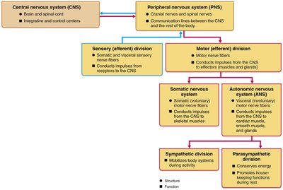
Sensory (Afferent) Division: Transmits sensory information from receptors to the CNS.
Motor (Efferent) Division: Transmits commands from the CNS to effector organs (muscles and glands).
Somatic Nervous System: Controls voluntary movements of skeletal muscles.
Autonomic Nervous System (ANS): Regulates involuntary functions (e.g., cardiac and smooth muscle, glands).
Sympathetic Division: Mobilizes body systems during activity (fight or flight).
Parasympathetic Division: Conserves energy and promotes housekeeping functions during rest.
Neuroglia and Neurons
Neuroglia (Glial Cells)
Neuroglia are supporting cells that protect, insulate, and nourish neurons. They are essential for the proper functioning of the nervous system.
Astrocytes: Support neurons, regulate the chemical environment, and assist in nutrient exchange.
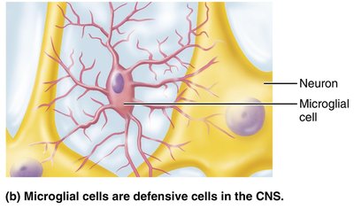
Microglial Cells: Act as immune defense cells in the CNS, phagocytizing debris and pathogens.
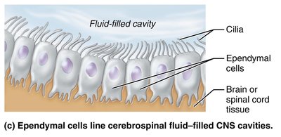
Ependymal Cells: Line cerebrospinal fluid-filled cavities and help circulate CSF.
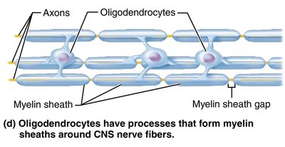
Oligodendrocytes: Form myelin sheaths around CNS nerve fibers.
Satellite Cells (PNS): Surround neuron cell bodies in ganglia, similar to astrocytes.
Schwann Cells (PNS): Form myelin sheaths around peripheral nerve fibers and assist in nerve regeneration.
Neurons (Nerve Cells)
Neurons are the excitable cells responsible for transmitting electrical signals throughout the nervous system. They have unique properties:
Extreme longevity: Can last a person's lifetime.
Amitotic: Most do not divide after development.
High metabolic rate: Require continuous oxygen and glucose.
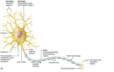
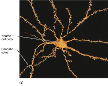
Structure: Consist of a cell body (soma), dendrites, and a single axon.
Neuron Processes
Dendrites: Short, branched processes that receive signals and convey them toward the cell body.
Axon: Long process that transmits impulses away from the cell body to other neurons or effectors.
Axon Terminals: Secretory regions that release neurotransmitters.
Myelin Sheath
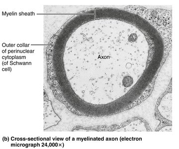
The myelin sheath is a white, fatty covering that insulates axons and increases the speed of impulse transmission.
In the PNS: Formed by Schwann cells wrapping around axons.
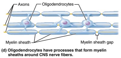
In the CNS: Formed by oligodendrocytes, each of which can myelinate multiple axons.
Nodes of Ranvier: Gaps between myelin segments where action potentials are regenerated.
Classification of Neurons
Structural Classification
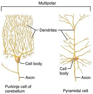
Multipolar Neurons: Many processes (1 axon, many dendrites); most common in CNS.
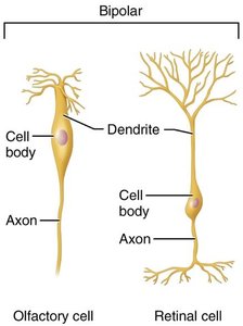
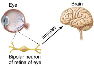
Bipolar Neurons: Two processes (1 axon, 1 dendrite); found in special sensory organs (retina, olfactory mucosa).
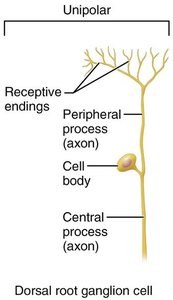
Unipolar (Pseudounipolar) Neurons: Single process that splits into two branches; mainly sensory neurons in PNS.
Functional Classification
Sensory (Afferent) Neurons: Transmit impulses from sensory receptors to the CNS; mostly unipolar.
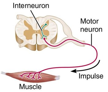
Motor (Efferent) Neurons: Carry impulses from the CNS to effectors; multipolar.
Interneurons (Association Neurons): Connect sensory and motor neurons within the CNS; most abundant type.
Membrane Potentials and Electrical Signaling
Resting Membrane Potential
Neurons maintain a resting membrane potential (typically around -70 mV) due to differences in ion concentrations and membrane permeability.
Key ions: Sodium (Na+), potassium (K+), chloride (Cl-), and negatively charged proteins.
Sodium-potassium pump: Maintains gradients by pumping 3 Na+ out and 2 K+ in.
Polarization: The inside of the neuron is more negative than the outside.
Changes in Membrane Potential
Depolarization: Membrane potential becomes less negative (closer to zero); increases likelihood of action potential.
Hyperpolarization: Membrane potential becomes more negative; decreases likelihood of action potential.
Graded Potentials
Graded potentials are short-lived, localized changes in membrane potential that decay with distance. They are essential for initiating action potentials.
Types: Receptor potentials, postsynaptic potentials, end-plate potentials.
Spread: Current flows locally and dissipates quickly.
Action Potentials
An action potential is a brief, large change in membrane potential that propagates along the axon without decrement. It is the primary means of long-distance neural communication.
Phases: Resting state, depolarization, repolarization, hyperpolarization.
All-or-none principle: An action potential either occurs fully or not at all.
Propagation: Action potentials travel in one direction along the axon.
Refractory periods: Absolute (no new AP possible) and relative (stronger stimulus needed).
Conduction Velocity
Factors affecting speed: Axon diameter (larger = faster), degree of myelination (myelinated = faster).
Types of fibers: A (fastest), B (intermediate), C (slowest).
Saltatory conduction: In myelinated axons, APs jump from node to node, increasing speed.
Synapses and Neurotransmission
Synapses
Synapses are specialized junctions where neurons communicate with other neurons or effector cells.
Electrical synapses: Direct, rapid communication via gap junctions; rare in adults.
Chemical synapses: Most common; use neurotransmitters to transmit signals across a synaptic cleft.
Steps in chemical synaptic transmission:
AP arrives at axon terminal.
Voltage-gated Ca2+ channels open; Ca2+ enters terminal.
Ca2+ triggers exocytosis of neurotransmitter vesicles.
Neurotransmitter diffuses across cleft and binds to postsynaptic receptors.
Ion channels open, generating graded potentials.
Neurotransmitter action is terminated by reuptake, degradation, or diffusion.
Postsynaptic Potentials
Excitatory Postsynaptic Potentials (EPSPs): Depolarize the postsynaptic membrane, increasing the chance of AP.
Inhibitory Postsynaptic Potentials (IPSPs): Hyperpolarize the membrane, decreasing the chance of AP.
Summation: EPSPs and IPSPs can summate temporally or spatially to influence AP generation.
Neurotransmitters
Types of Neurotransmitters
Acetylcholine (ACh): First discovered; used at neuromuscular junctions and in the CNS and PNS.
Biogenic Amines: Dopamine, norepinephrine, epinephrine, serotonin, histamine; involved in mood and behavior.
Amino Acids: Glutamate, aspartate, glycine, GABA.
Peptides: Substance P, endorphins, somatostatin, cholecystokinin.
Purines: ATP, adenosine.
Gases and Lipids: Nitric oxide, carbon monoxide, endocannabinoids.
Neurotransmitter Actions
Excitatory vs. Inhibitory: Some neurotransmitters can have different effects depending on the receptor type.
Direct vs. Indirect: Direct neurotransmitters open ion channels; indirect act through second messengers (e.g., G protein-coupled receptors).
Neuromodulators: Chemicals that modulate synaptic transmission without directly causing EPSPs or IPSPs.
Neural Integration and Circuits
Neural Processing
Serial Processing: Information travels in a single pathway (e.g., reflex arcs).
Parallel Processing: Information is processed simultaneously along multiple pathways, allowing complex responses.
Types of Neural Circuits
Diverging: One input, many outputs (amplifies signal).
Converging: Many inputs, one output (concentrates signal).
Reverberating: Signal travels through a chain of neurons, each feeding back to previous neurons (oscillations, rhythmic activity).
Parallel After-Discharge: Signal stimulates several neurons in parallel arrays (complex processing).
Developmental Aspects of Neurons
The nervous system develops from the neural tube and neural crest. Neurons are generated, migrate, and form synaptic connections. Synaptic pruning and plasticity are essential for learning and memory, especially during childhood.
*Additional info: This guide expands on the provided notes with definitions, examples, and context to ensure a comprehensive, self-contained study resource for college-level Anatomy & Physiology students.*
