 Back
BackFundamentals of the Nervous System: Structure, Function, and Electrical Properties
Study Guide - Smart Notes
 Tailored notes based on your materials, expanded with key definitions, examples, and context.
Nervous System Fundamentals
Functions of the Nervous System
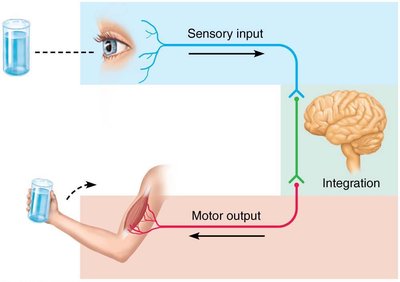
The nervous system is the master controlling and communicating system of the body. It operates through electrical and chemical signals, enabling rapid and precise communication. The nervous system performs three main functions:
Sensory Input: Information is gathered by sensory receptors about internal and external changes.
Integration: The processing and interpretation of sensory input occurs in the central nervous system.
Motor Output: Activation of effector organs (muscles and glands) produces a response.
Divisions of the Nervous System
Central and Peripheral Nervous Systems
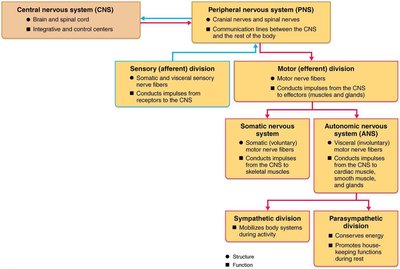
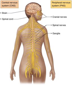
The nervous system is divided into two main parts:
Central Nervous System (CNS): Consists of the brain and spinal cord, located in the dorsal body cavity. It serves as the integration and control center.
Peripheral Nervous System (PNS): Composed of nerves that extend from the brain and spinal cord. The PNS is further divided into:
Sensory (afferent) division: Transmits impulses to the CNS from sensory receptors.
Motor (efferent) division: Transmits impulses from the CNS to effector organs. This includes:
Somatic nervous system: Voluntary control of skeletal muscles.
Autonomic nervous system (ANS): Involuntary control of smooth muscle, cardiac muscle, and glands. The ANS is subdivided into sympathetic (mobilizes body systems) and parasympathetic (conserves energy) divisions.
Neuroglia (Glial Cells)
Types and Functions of Neuroglia
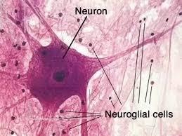
Nervous tissue contains two principal cell types: neuroglia (glial cells) and neurons. Neuroglia are small cells that support, protect, and insulate neurons.
Astrocytes: Most abundant CNS glial cells; support neurons, regulate exchanges between capillaries and neurons, and control the chemical environment.
Microglial cells: Defensive cells in the CNS; monitor neurons and can transform to phagocytize microorganisms and debris.
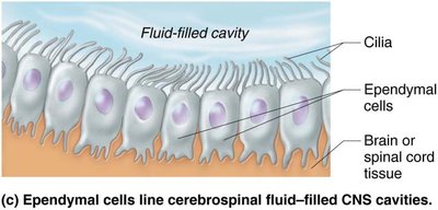
Ependymal cells: Line cerebrospinal fluid-filled CNS cavities; may be ciliated to circulate CSF.
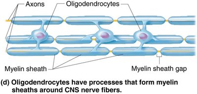
Oligodendrocytes: Form myelin sheaths around CNS nerve fibers.
Satellite cells (PNS): Surround neuron cell bodies in the PNS; function similar to astrocytes.
Schwann cells (PNS): Form myelin sheaths in the PNS; vital for regeneration of damaged peripheral nerve fibers.


Neurons: Structure and Function
Neuron Anatomy
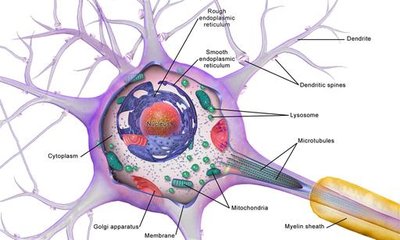
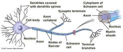
Neurons are the structural units of the nervous system, specialized for conducting impulses. They exhibit extreme longevity, are mostly amitotic, and have a high metabolic rate. Each neuron consists of a cell body (soma) and one or more processes (dendrites and axons).
Cell Body (Soma): Biosynthetic center; contains organelles for protein synthesis and metabolic activity.
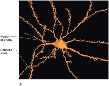
Dendrites: Receptive regions; convey incoming messages toward the cell body as graded potentials.
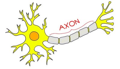
Axon: Conducting region; generates and transmits nerve impulses away from the cell body.


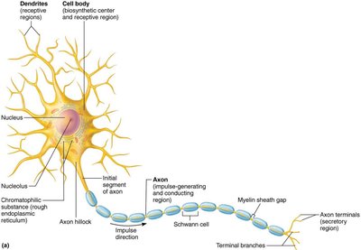
Myelination
Myelin is a whitish, protein-lipid substance that insulates axons and increases the speed of impulse transmission.
Myelinated fibers: Conduct impulses rapidly.
Nonmyelinated fibers: Conduct impulses slowly.
Myelination in PNS: Schwann cells wrap around axons, forming myelin sheaths and nodes of Ranvier.
Myelination in CNS: Oligodendrocytes form myelin sheaths; no outer collar of perinuclear cytoplasm.

Classification of Neurons
Structural Classification
Neurons are classified by the number of processes extending from the cell body:
Multipolar: Three or more processes (one axon, multiple dendrites); most common in CNS.
Bipolar: Two processes (one axon, one dendrite); found in special sensory organs.
Unipolar (pseudounipolar): One T-like process; found in sensory neurons of PNS.

Functional Classification
Neurons are also classified by the direction of impulse transmission:
Sensory (afferent): Transmit impulses toward CNS; mostly unipolar.
Motor (efferent): Carry impulses from CNS to effectors; multipolar.
Interneurons: Lie between motor and sensory neurons; multipolar, mostly in CNS.
Basic Principles of Electricity in Neurons
Electrical Properties
Neurons operate based on electrical principles:
Voltage: Potential energy generated by separated charge; measured in volts (V).
Current: Flow of electrical charge (ions) between two points.
Resistance: Hindrance to charge flow; insulators have high resistance, conductors have low resistance.
Ohm’s Law:
Membrane Ion Channels
Ion channels are large proteins that allow selective ion movement across the membrane. Types include:
Leakage channels: Always open.
Gated channels: Open/close in response to stimuli (chemical, voltage, mechanical).
Electrochemical Gradient
The combined electrical and chemical gradients drive ion movement, creating electrical currents and voltage changes across the membrane.
Resting Membrane Potential
Generation and Maintenance
The resting membrane potential of a neuron is approximately −70 mV, with the inside of the cell being more negative than the outside. This is established by:
Differences in ionic composition: ECF has higher Na+, ICF has higher K+.
Differences in membrane permeability: More K+ diffuses out than Na+ diffuses in.
Sodium-potassium pump: Maintains gradients by pumping 3 Na+ out and 2 K+ in.
Changes in Membrane Potential
Types of Signals
Graded potentials: Short-lived, localized changes; operate over short distances.
Action potentials: Long-distance signals; brief reversal of membrane potential (~100 mV).
Membrane Potential Changes
Depolarization: Membrane potential moves toward zero; increases probability of impulse.
Hyperpolarization: Membrane potential moves away from zero; decreases probability of impulse.
Action Potential Generation
Steps of Action Potential
Resting state: All gated Na+ and K+ channels closed; only leakage channels open.
Depolarization: Na+ channels open; Na+ influx causes membrane potential to rise.
Repolarization: Na+ channels inactivate, K+ channels open; K+ efflux restores negative potential.
Hyperpolarization: Some K+ channels remain open; membrane potential dips below resting.
Propagation and Coding
Propagation: AP travels down axon; self-propagating.
Stimulus Intensity: Coded by frequency of APs, not size.
Refractory Periods
Absolute refractory period: No new AP can be generated.
Relative refractory period: AP can be generated only by strong stimulus.
Conduction Velocity
Axon diameter: Larger diameter = faster conduction.
Degree of myelination: Myelinated axons conduct faster (saltatory conduction).
Clinical Relevance: Multiple Sclerosis
Pathophysiology and Symptoms
Multiple sclerosis (MS) is an autoimmune disease where myelin sheaths in the CNS are destroyed, leading to impaired impulse conduction. Symptoms include visual disturbances, weakness, loss of muscular control, speech disturbances, and incontinence. 
The Synapse
Types and Function
Synapses are junctions that mediate information transfer between neurons.
Electrical synapses: Neurons are electrically coupled via gap junctions; rapid communication.
Chemical synapses: Specialized for release and reception of neurotransmitters; involve synaptic cleft.
Chemical Synapse Transmission Steps
AP arrives at axon terminal.
Voltage-gated Ca2+ channels open; Ca2+ enters terminal.
Ca2+ causes synaptic vesicles to release neurotransmitter by exocytosis.
Neurotransmitter diffuses across synaptic cleft and binds to receptors on postsynaptic membrane.
Binding opens ion channels, creating graded potentials.
Neurotransmitter effects terminated by reuptake, degradation, or diffusion.
Postsynaptic Potentials and Integration
Types of Postsynaptic Potentials
EPSP (excitatory): Depolarizes postsynaptic membrane; increases likelihood of AP.
IPSP (inhibitory): Hyperpolarizes postsynaptic membrane; decreases likelihood of AP.
Summation and Potentiation
Temporal summation: Rapid-fire impulses from one neuron.
Spatial summation: Simultaneous stimulation by multiple neurons.
Synaptic potentiation: Repeated use increases synaptic strength; important for learning and memory.
Neurotransmitters
Classification by Structure
Acetylcholine (ACh): Used at neuromuscular junctions and by many CNS/ANS neurons.
Biogenic amines: Dopamine, norepinephrine, epinephrine, serotonin; roles in emotion and biological clock.
Amino acids: Glutamate, aspartate, glycine, GABA.
Peptides: Substance P, endorphins, gut-brain peptides.
Purines: ATP, adenosine.
Gasotransmitters: Nitric oxide (NO), endocannabinoids.
Classification by Function
Effects: Excitatory (depolarizing) or inhibitory (hyperpolarizing); effect depends on receptor.
Actions: Direct (binds and opens ion channels) or indirect (acts through second messengers).
Neural Integration
Neuronal Pools and Processing
Neurons function in groups called neuronal pools, integrating incoming information and forwarding processed information.
Serial processing: Input travels along one pathway; produces specific response (e.g., reflexes).
Parallel processing: Input travels along several pathways; important for higher-level functions.
Types of Circuits
Four main types of circuits exist in neuronal pools, each with distinct patterns of synaptic connections.
Summary Table: Structural Classes of Neurons
Type | Processes | Location | Function |
|---|---|---|---|
Multipolar | 3+ (1 axon, others dendrites) | CNS | Motor, interneurons |
Bipolar | 2 (1 axon, 1 dendrite) | Special sensory organs | Sensory |
Unipolar | 1 T-like process | PNS | Sensory |
