 Back
BackGross Anatomy of the Brain and Cranial Nerves: Structured Study Notes
Study Guide - Smart Notes
 Tailored notes based on your materials, expanded with key definitions, examples, and context.
Gross Anatomy of the Brain and Cranial Nerves
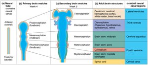
Embryonic Development and Brain Regions
The human brain develops from the neural tube, which forms primary and secondary brain vesicles. These vesicles give rise to the major adult brain structures and associated neural canal regions.
Neural Tube: The precursor to the central nervous system, containing the neural canal.
Primary Brain Vesicles: Prosencephalon (forebrain), Mesencephalon (midbrain), Rhombencephalon (hindbrain).
Secondary Brain Vesicles: Telencephalon, Diencephalon, Mesencephalon, Metencephalon, Myelencephalon.
Adult Brain Structures: Cerebrum, diencephalon, brain stem (midbrain, pons, medulla oblongata), cerebellum.
Neural Canal Regions: Lateral ventricles, third ventricle, cerebral aqueduct, fourth ventricle, central canal.
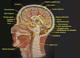
Major Brain Structures: Sagittal View
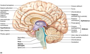
The sagittal view of the brain reveals key anatomical features, including the cerebrum, cerebellum, brainstem, and associated ventricles. Understanding these structures is essential for identifying their functions and relationships.
Cerebrum: Responsible for higher cognitive functions.
Corpus Callosum: Connects the two cerebral hemispheres.
Thalamus: Relay center for sensory information.
Hypothalamus: Regulates homeostasis and endocrine functions.
Pituitary Gland: Major endocrine gland.
Pons and Medulla Oblongata: Control vital autonomic functions.
Cerebellum: Coordinates movement and balance.
Ventricles: Spaces containing cerebrospinal fluid (CSF).
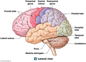
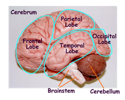
Cerebral Lobes and Surface Features
The cerebrum is divided into lobes, each associated with specific functions. Surface features include gyri (ridges), sulci (grooves), and fissures (deep grooves).
Frontal Lobe: Motor control, problem-solving, speech production.
Parietal Lobe: Sensory processing, spatial orientation.
Temporal Lobe: Auditory processing, memory.
Occipital Lobe: Visual processing.
Insula: Deep within the lateral sulcus, involved in taste and visceral sensation.
Gyri: Elevated ridges on the cerebral surface.
Sulci: Shallow grooves separating gyri.
Fissures: Deep grooves dividing major brain regions.
Gray Matter and White Matter
The brain is composed of gray matter and white matter, each with distinct functions and locations.
Gray Matter: Contains neuronal cell bodies; forms the cerebral cortex and deep nuclei (e.g., thalamus, hypothalamus).
White Matter: Composed of myelinated axons; includes structures like the corpus callosum and arbor vitae of the cerebellum.
Cerebral Cortex: Surface layer of gray matter responsible for higher-order functions.
Nuclei: Clusters of gray matter deep within the brain.
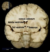
Diencephalon and Brainstem
The diencephalon and brainstem are central to sensory processing, autonomic regulation, and relay of information between brain regions.
Diencephalon: Includes the thalamus, hypothalamus, pineal gland, and associated structures.
Thalamus: Sensory relay station.
Hypothalamus: Controls autonomic and endocrine functions.
Pineal Gland: Secretes melatonin, regulates sleep-wake cycles.
Brainstem: Composed of midbrain, pons, and medulla oblongata; controls vital functions.
Cerebral Aqueduct: Connects third and fourth ventricles.
Meninges and Cerebrospinal Fluid (CSF)
The brain is protected by three meningeal layers and bathed in cerebrospinal fluid, which cushions and nourishes neural tissue.
Dura Mater: Outermost, tough layer; has periosteal and meningeal components.
Arachnoid Mater: Middle layer; contains subarachnoid space filled with CSF.
Pia Mater: Innermost, delicate layer; adheres to brain surface.
CSF Production: Occurs in choroid plexuses within ventricles.
CSF Flow: Lateral ventricles → interventricular foramen → third ventricle → cerebral aqueduct → fourth ventricle → central canal or subarachnoid space.
Arachnoid Villi: Drain CSF into dural sinuses, especially the superior sagittal sinus.
Cranial Nerves
The twelve cranial nerves are identified by name and number, each with specific sensory, motor, or mixed functions.
Number | Name | Main Function |
|---|---|---|
I | Olfactory | Smell |
II | Optic | Vision |
III | Oculomotor | Eye movement |
IV | Trochlear | Eye movement |
V | Trigeminal | Facial sensation |
VI | Abducens | Eye movement |
VII | Facial | Facial movement |
VIII | Auditory (Vestibulocochlear) | Hearing and balance |
IX | Glossopharyngeal | Organs and taste |
X | Vagus | Organs and taste |
XI | Accessory | Shoulder shrug & head turn |
XII | Hypoglossal | Tongue movement |
Summary Table: Brain Regions and Functions
Region | Main Function |
|---|---|
Frontal Lobe | Motor control, reasoning, speech |
Parietal Lobe | Sensory processing, spatial awareness |
Temporal Lobe | Auditory processing, memory |
Occipital Lobe | Visual processing |
Insula | Taste, visceral sensation |
Thalamus | Sensory relay |
Hypothalamus | Homeostasis, endocrine regulation |
Cerebellum | Coordination, balance |
Brainstem | Autonomic functions |
Example: The occipital lobe is primarily involved in visual processing, receiving and interpreting information from the eyes.
Additional info: The notes above expand on brief points from the original material, providing definitions, context, and logical grouping for clarity and completeness.
