 Back
BackHistology and Tissue Organization: Study Guide for Anatomy & Physiology
Study Guide - Smart Notes
 Tailored notes based on your materials, expanded with key definitions, examples, and context.
Histology: The Study of Tissues
Introduction to Tissues
Histology is the branch of anatomy that studies tissues, which are groups of cells with a common function. Tissues join at cellular junctions and collectively form organs, which in turn make up body systems. There are four primary tissue types in the human body: epithelial, connective, muscular, and nervous.
Tissue: Group of cells with a common function, joined at their plasma membranes.
Organ: Structure composed of multiple tissue types working together.
Histology: The microscopic study of tissues.
Classification of Tissues
Main Tissue Types and Their Subtypes
Epithelial Tissue: Covers surfaces, lines cavities, and forms glands.
Connective Tissue: Supports, binds, and protects organs; most abundant tissue type.
Muscular Tissue: Responsible for movement.
Nervous Tissue: Initiates and transmits electrical impulses.
Epithelial Tissue
Characteristics and Classification
Epithelial tissue forms the covering or lining of all internal and external body surfaces. It is avascular (lacks blood vessels), receives nutrients from underlying connective tissue, and is separated from connective tissue by a basement membrane. Epithelial cells are tightly packed and have a high rate of mitosis.
Functions: Protection, absorption, secretion, excretion, sensation, and filtration.
Location: Epidermis, lining of hollow organs, glands.
Basement Membrane: Separates epithelial from connective tissue; has apical (top) and basal (bottom) regions.
Types of Epithelial Tissue
Simple Epithelium: One cell layer thick; all cells touch the basement membrane.
Stratified Epithelium: Two or more layers; only the deepest layer touches the basement membrane.
Cell shapes include:
Squamous: Flat, scale-like
Cuboidal: Cube-shaped, equal sides
Columnar: Taller than wide, may have cilia or microvilli
Discoid: Disc-shaped (e.g., red blood cells)
Major Types of Epithelial Tissue
Simple Squamous Epithelium
Simple Cuboidal Epithelium
Simple Columnar Epithelium
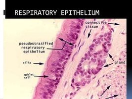
Pseudostratified Ciliated Columnar Epithelium
Stratified Squamous Epithelium (keratinized and nonkeratinized)
Stratified Cuboidal Epithelium
Stratified Columnar Epithelium
Transitional Epithelium
Specialized Structures
Cilia: Move substances across the surface (e.g., trachea).
Microvilli: Increase surface area for absorption (e.g., intestines).
Goblet Cells: Secrete mucus, found in columnar epithelium.
Example: Pseudostratified Ciliated Columnar Epithelium
This tissue appears to have multiple layers due to varying cell heights but is actually a single layer with all cells touching the basement membrane. It is found in the respiratory tract, where cilia move mucus and trapped particles.
Location: Trachea, upper respiratory tract
Function: Secretion and movement of mucus
Connective Tissue
Overview and Functions
Connective tissue is the most abundant tissue type, found throughout the body. It supports, binds, and protects organs, and is characterized by cells scattered within an extracellular matrix. Most connective tissues are vascular, except cartilage, tendons, and ligaments.
Functions: Binding, support, protection, movement, storage, transport, heat production.
Matrix: Composed of fibers (collagen, elastic, reticular) and ground substance (mainly water).
Cells: Fibroblasts, adipocytes, osteocytes, chondrocytes, blood cells.
Types of Connective Tissue
Loose Connective Tissue: Areolar, adipose
Dense Connective Tissue: Tendons, ligaments
Cartilage: Hyaline, elastic, fibrocartilage
Bone: Compact (osteons), spongy (trabeculae)
Blood and Lymph: Only fluid connective tissues
Connective Tissue Cell Types
-blast: Immature, matrix-producing cell (e.g., osteoblast)
-cyte: Mature cell, maintains matrix (e.g., chondrocyte)
-clast: Breaks down matrix (e.g., osteoclast)
Muscular Tissue
Types and Characteristics
Muscular tissue is specialized for contraction and movement. There are three types, each with distinct features and functions.
Feature | Skeletal | Cardiac | Smooth |
|---|---|---|---|
Location | Attached to bones | Heart | Walls of hollow organs |
Control | Voluntary | Involuntary | Involuntary |
Striations | Yes | Yes | No |
Intercalated Discs | No | Yes | No |
Nuclei | Multiple, peripheral | Single, central | Single, central |
Additional info: Skeletal muscles are named based on fiber direction, size, number of origins, location, shape, and action.
Nervous Tissue
Structure and Function
Nervous tissue is specialized for communication via electrical impulses. It is found in the brain, spinal cord, and nerves. There are two main cell types:
Neurons: Transmit impulses; classified by function (sensory, interneuron, motor) and structure (unipolar, bipolar, multipolar).
Neuroglia (Glial Cells): Support, protect, and nourish neurons; more abundant than neurons.
Integumentary System: The Skin
Structure of the Skin
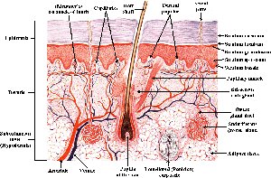
The skin is the largest organ of the body and consists of two main layers: the epidermis and the dermis. The epidermis is made of stratified squamous keratinized epithelium, while the dermis is connective tissue. Associated structures include hair, nails, and glands.
Epidermis: Stratified squamous epithelium; avascular; provides protection.
Dermis: Connective tissue; contains blood vessels, nerves, glands, and hair follicles.
Basement Membrane: Separates epidermis from dermis.
Burns and Skin Injury
First-degree: Only epidermis affected.
Second-degree: Epidermis and part of dermis affected.
Third-degree: Extends into deeper tissues; may require grafting.
Dehydration is a major concern in burn patients; treatment often involves IV fluids.
Skin Cancer
Basal Cell Carcinoma: Most common, least dangerous.
Melanoma: Most dangerous, high risk of metastasis.
Assessment (A, B, C, D Rule): Asymmetry, Border, Color, Diameter.
Wound Healing and Inflammation
Steps of Wound Healing
Hemostasis: Stopping bleeding.
Inflammation: White blood cells clean the site.
Proliferation: New tissue and blood vessels form.
Remodeling: Wound closes; tissue matures (regenerative or fibrotic).
Cardinal Signs of Inflammation
Pain
Heat
Redness
Swelling
Loss of function
Key Terms and Concepts
Mutation: Change in DNA sequence
Phenotype: Observable traits
Genotype: Genetic makeup
Benign vs. Malignant: Non-cancerous vs. cancerous growth
Metastasis: Spread of cancer cells
Apoptosis: Programmed cell death
Necrosis: Unplanned cell death due to injury
