 Back
BackHistology: The Study of Tissues – Structure, Function, and Classification
Study Guide - Smart Notes
 Tailored notes based on your materials, expanded with key definitions, examples, and context.
Histology: The Study of Tissues
Introduction to Tissues
Histology is the branch of anatomy that studies tissues, which are groups of cells working together to perform specific functions. Understanding tissues is fundamental to comprehending how organs and organ systems operate in the human body. There are four primary tissue types: epithelial, connective, muscular, and nervous.
Tissue: A group of cells with a common function.
Organ: A structure composed of two or more tissue types working together for a specific function.
Cell Shapes and Sizes in Tissues
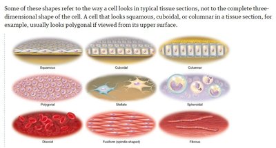
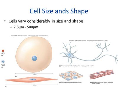
Cells in tissues exhibit a variety of shapes and sizes, which are crucial for their specific functions. The shape of a cell in a tissue section may differ from its three-dimensional form.
Common cell shapes: Squamous (flat), cuboidal (cube-shaped), columnar (tall), polygonal, stellate (star-shaped), spheroidal, discoid, fusiform (spindle-shaped), and fibrous.
Cell size: Ranges from 7.5 μm to 500 μm.
Overview of the Four Primary Tissue Types
Tissue Type | Main Functions | Key Features |
|---|---|---|
Epithelial | Protection, secretion, absorption, excretion, filtration, sensation | Cells closely packed, avascular, polar (apical and basal surfaces) |
Connective | Binding, support, protection, movement, storage, heat production, transport | Most abundant, diverse, vascular (except cartilage), cells + matrix |
Muscular | Movement, posture, heat production | Elongated cells (fibers), contractile proteins |
Nervous | Rapid communication, control, integration | Neurons and supporting glial cells |
Epithelial Tissue
Characteristics and Classification
Epithelial tissue covers body surfaces, lines cavities, and forms glands. It is characterized by closely packed cells with minimal extracellular material, polarity (distinct apical and basal surfaces), and avascularity (lacks blood vessels; nutrients diffuse from underlying connective tissue).
Polarity: Apical surface (exposed), basal surface (attached to basement membrane).
Basement membrane: Thin layer separating epithelium from connective tissue below.
Avascular: No direct blood supply; relies on diffusion from connective tissue.
Types of Epithelial Tissue
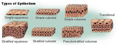
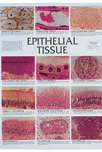
Epithelial tissues are classified by the number of cell layers and cell shape:
Simple epithelium: One cell layer; all cells touch the basement membrane.
Stratified epithelium: Two or more layers; only the deepest layer touches the basement membrane.
Pseudostratified epithelium: Appears layered but all cells contact the basement membrane.
Transitional epithelium: Specialized for stretching (e.g., urinary bladder).
Common Epithelial Types and Locations
Type | Location | Function |
|---|---|---|
Simple squamous | Alveoli, blood vessels (endothelium) | Diffusion, filtration |
Simple cuboidal | Kidney tubules, glands | Secretion, absorption |
Simple columnar | Digestive tract lining | Absorption, secretion |
Pseudostratified ciliated columnar | Respiratory tract | Secretion, movement of mucus |
Stratified squamous (keratinized) | Epidermis of skin | Protection (dry areas) |
Stratified squamous (nonkeratinized) | Mouth, esophagus, vagina | Protection (moist areas) |
Transitional | Urinary bladder | Stretching, distension |
Functions of Epithelial Tissue
Protection: Shields underlying tissues from mechanical and chemical stress.
Secretion: Forms glands that release hormones, enzymes, and other substances.
Absorption: Uptake of nutrients and fluids (e.g., intestines).
Excretion: Removal of waste products.
Filtration: Selective movement of substances (e.g., kidneys).
Sensation: Contains nerve endings for sensory reception.
Specialized Epithelial Structures
Cilia: Hair-like projections for movement of substances (e.g., respiratory tract).
Microvilli: Increase surface area for absorption (e.g., intestines).
Goblet cells: Unicellular glands that secrete mucus.
Glandular Epithelium
Exocrine glands: Secrete products into ducts or onto surfaces (e.g., sweat, salivary glands).
Endocrine glands: Ductless; secrete hormones directly into the bloodstream (e.g., thyroid, adrenal glands).
Mixed glands: Both exocrine and endocrine functions (e.g., pancreas, gonads).
Connective Tissue
Characteristics and Classification
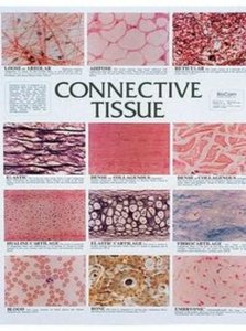
Connective tissue is the most abundant and diverse tissue type in the body. It supports, binds, and protects organs. All connective tissues consist of cells and an extracellular matrix (fibers and ground substance).
Vascularity: Most are vascular (except cartilage).
Matrix: Non-living material between cells, composed of fibers (collagen, elastic, reticular) and ground substance (mainly water).
Origin: All connective tissues arise from embryonic mesenchyme (mesoderm).
Major Types of Connective Tissue
Type | Subtypes | Location | Function |
|---|---|---|---|
Cartilage | Hyaline (most abundant), elastic, fibrocartilage | Joints, ear, intervertebral discs | Support, flexibility, cushioning |
Bone (Osseous) | Compact, spongy | Skeletal system | Support, protection, movement |
Blood | Only fluid connective tissue | Blood vessels | Transport, immunity |
Dense connective | Regular (tendons, ligaments), irregular | Tendons, ligaments, dermis | Binding, strength |
Loose connective | Areolar, adipose, reticular | Under skin, around organs | Cushioning, energy storage |
Cells of Connective Tissue
-blast: Immature, matrix-producing cells (e.g., fibroblast, chondroblast, osteoblast).
-cyte: Mature cells maintaining the tissue (e.g., chondrocyte, osteocyte).
-clast: Cells involved in breakdown (e.g., osteoclast).
Functions of Connective Tissue
Binding and support (e.g., tendons, ligaments)
Protection (e.g., bone, cartilage)
Immune defense (e.g., blood, lymphatic tissue)
Movement (e.g., bones as levers)
Storage (e.g., adipose tissue stores fat)
Heat production (e.g., brown fat)
Transport (e.g., blood)
Muscular Tissue
Types of Muscle Tissue
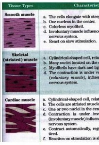
Muscle tissue is specialized for contraction and movement. There are three types:
Skeletal muscle: Voluntary, striated, multinucleated, attached to bones.
Cardiac muscle: Involuntary, striated, single nucleus, intercalated discs, found in the heart.
Smooth muscle: Involuntary, non-striated, single nucleus, found in walls of hollow organs.
Nervous Tissue
Structure and Function
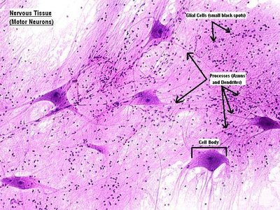
Nervous tissue is specialized for communication by electrical and chemical signals. It consists of neurons (nerve cells) and supporting glial cells.
Neurons: Transmit impulses; have cell body, axon, and dendrites.
Glial cells: Support, protect, and nourish neurons.
Cell Junctions
Types and Locations
Tight junctions: Seal cells together; found in stomach and intestines.
Gap junctions: Allow communication; found in heart muscle (intercalated discs).
Desmosomes: Anchor cells; found in epidermis and cardiac muscle.
Embryonic Origin of Tissues
Ectoderm: Forms epidermis and nervous system.
Mesoderm: Forms mesenchyme, which gives rise to connective tissues, muscle, and blood.
Endoderm: Forms digestive glands and mucous membranes of digestive and respiratory tracts.
Tissue Repair and Pathology
Repair Mechanisms
Regeneration: Replacement of dead/damaged cells with original cells (minor injuries).
Fibrosis: Replacement with scar tissue (major injuries); does not restore normal function.
Stages of Wound Healing
Tissue injury with bleeding
Formation of blood clot (inflammation, homeostasis)
Proliferation (granulation tissue forms)
Maturation (remodeling and strengthening)
Key Terms
Atrophy: Decrease in size of tissue or organ.
Necrosis: Sudden tissue death.
Apoptosis: Programmed cell death.
Gangrene: Tissue necrosis due to insufficient blood supply.
Decubitus ulcer: Pressure sore from prolonged immobility.
Infarction: Sudden tissue death due to loss of blood supply (e.g., myocardial infarction).
Summary Table: Epithelial vs. Connective Tissue
Feature | Epithelial Tissue | Connective Tissue |
|---|---|---|
Cell arrangement | Closely packed, little matrix | Widely spaced, much matrix |
Vascularity | Avascular | Vascular (except cartilage) |
Location | Surfaces, linings, glands | Throughout body, under epithelia |
Functions | Protection, secretion, absorption | Support, binding, transport |
Additional info: For further study, refer to textbook Table 5.1 for a comprehensive overview of tissue types, and review histological slides for visual identification of tissues in laboratory settings.
