 Back
BackHuman Reflex Physiology: Structure and Function of Reflex Arcs
Study Guide - Smart Notes
 Tailored notes based on your materials, expanded with key definitions, examples, and context.
Reflexes and Reflex Arcs
Definition and Overview
Reflexes are rapid, involuntary responses to specific stimuli that help maintain homeostasis and protect the body from harm. These responses can be either somatic (involving skeletal muscles) or autonomic (involving smooth muscle, cardiac muscle, or glands). Reflex arcs are the neural pathways that mediate these reflex actions, allowing for quick and automatic responses without the need for conscious thought.
Reflex: An automatic, involuntary response to a stimulus.
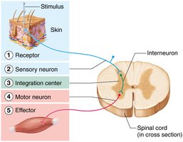
Reflex arc: The neural pathway that mediates a reflex, typically involving a receptor, sensory neuron, integration center, motor neuron, and effector.
Importance: Reflex testing is a key part of physical examinations, as abnormal reflexes can indicate neurological dysfunction.

The Reflex Arc: Structure and Components
Pathway of a Reflex Arc
The reflex arc is the basic functional unit of the nervous system responsible for producing reflex actions. It consists of five main components:
Receptor: Detects the stimulus (e.g., pain, stretch, temperature).
Sensory neuron: Transmits the afferent impulse to the central nervous system (CNS).
Integration center: Usually one or more interneurons in the CNS (spinal cord or brain) that process the information.
Motor neuron: Conducts efferent impulses from the integration center to an effector.
Effector: Muscle or gland that responds to the motor neuron impulse.
Types of Reflexes
Somatic vs. Autonomic (Visceral) Reflexes
Reflexes can be classified based on the effectors they activate:
Somatic reflexes: Involve skeletal muscles (e.g., patellar reflex, corneal reflex).
Autonomic (visceral) reflexes: Involve smooth muscle, cardiac muscle, or glands (e.g., photopupillary reflex, salivary reflex).
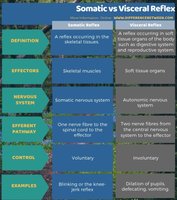
Feature | Somatic Reflex | Visceral (Autonomic) Reflex |
|---|---|---|
Definition | Reflex in skeletal tissues | Reflex in soft tissue organs |
Effectors | Skeletal muscles | Soft tissue organs |
Nervous System | Somatic nervous system | Autonomic nervous system |
Control | Voluntary | Involuntary |
Examples | Knee-jerk reflex | Pupil dilation, defecation |
Cranial vs. Spinal Reflexes
Spinal reflexes: Integrated in the spinal cord (e.g., withdrawal from pain, patellar reflex).
Cranial reflexes: Integrated in the brainstem (e.g., corneal reflex, gag reflex).
Ipsilateral, Contralateral, and Consensual Reflexes
Ipsilateral reflex: Response occurs on the same side as the stimulus.
Contralateral reflex: Response occurs on the opposite side of the stimulus.
Consensual reflex: Reflex observed on one side of the body when the other side is stimulated (e.g., pupillary light reflex).
Examples of Human Reflexes
Stretch Reflexes
Stretch reflexes are monosynaptic, spinal, and somatic. They help maintain muscle tone and posture by causing muscle contraction in response to stretching within the muscle.
Receptors: Muscle spindles (in muscles), Golgi tendon organs (in tendons).
Examples: Patellar reflex, Achilles reflex, biceps reflex.
Patellar Reflex (Knee-Jerk Reflex)
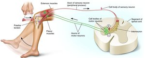
The patellar reflex is a classic example of a monosynaptic stretch reflex. Tapping the patellar tendon stretches the quadriceps muscle, triggering contraction and extension of the lower leg.
Type: Somatic, spinal reflex
Pathway: Involves L2-L4 spinal nerves
Receptors: Golgi tendon organs in the patellar ligament
Response: Extension of the lower leg
Plantar Reflex
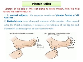
The plantar reflex is a polysynaptic, somatic, and spinal reflex. Stroking the lateral aspect of the sole of the foot normally causes flexion of the toes. An abnormal response (Babinski sign) is dorsiflexion of the big toe and fanning of the other toes, which may indicate neurological damage in adults.
Type: Somatic, spinal reflex
Pathway: Involves L5, S1 spinal nerves
Normal response: Flexion of toes
Abnormal response: Babinski sign
Gag Reflex
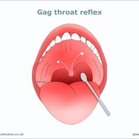
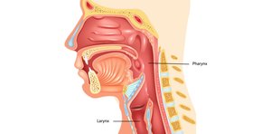
The gag reflex is a protective somatic and cranial reflex that prevents foreign objects from entering the throat. Stimulation of the posterior pharynx triggers contraction of the pharyngeal muscles, resulting in gagging.
Type: Somatic, cranial reflex
Pathway: Involves cranial nerve IX (glossopharyngeal nerve)
Response: Contraction of pharyngeal muscles (gagging)
Photopupillary Reflex
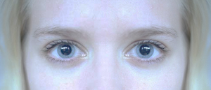
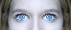
The photopupillary reflex is an autonomic, cranial, and consensual reflex. Exposure of one eye to bright light causes constriction of both pupils, protecting the retina from excessive light.
Type: Autonomic, cranial, consensual reflex
Pathway: Involves cranial nerve III (oculomotor nerve)
Response: Constriction of both pupils
Summary Table: Key Reflexes
Reflex | Type | Integration Center | Effector | Normal Response |
|---|---|---|---|---|
Patellar | Somatic, spinal | Spinal cord (L2-L4) | Quadriceps | Leg extension |
Achilles | Somatic, spinal | Spinal cord (S1-S2) | Gastrocnemius, soleus | Plantar flexion |
Plantar | Somatic, spinal | Spinal cord (L5, S1) | Toe flexors | Toe flexion |
Gag | Somatic, cranial | Brainstem | Pharyngeal muscles | Gagging |
Photopupillary | Autonomic, cranial | Brainstem | Pupillary muscles | Pupil constriction |
Additional info:
Monosynaptic reflexes involve a single synapse between a sensory and a motor neuron (e.g., patellar reflex).
Polysynaptic reflexes involve one or more interneurons (e.g., withdrawal reflex, plantar reflex).
Reflex testing is used clinically to assess the integrity of the nervous system at various levels.
