 Back
BackImmune, Respiratory, and Digestive Systems: Structured Study Notes
Study Guide - Smart Notes
 Tailored notes based on your materials, expanded with key definitions, examples, and context.
Immune System
Overview of the Immune System
The immune system provides resistance to disease by defending the body against pathogens such as bacteria, fungi, and viruses. Unlike anatomical organ systems, it is a functional system composed of immune cells and molecules that reside in lymphoid tissues and circulate in body fluids. The immune system is divided into two main subsystems: the innate (nonspecific) defense system and the adaptive (specific) defense system.
Innate defense system: First and second lines of defense; responds quickly and non-specifically.
Adaptive defense system: Third line of defense; attacks particular foreign substances and takes longer to react.
Innate Immune System: First Line of Defense
The first line of defense consists of surface membranes (skin and mucous membranes) and their secretions, which act as physical and chemical barriers to most microorganisms.
Acid: Acidity of skin and mucous secretions inhibits microbial growth (acid mantle).
Enzymes: Lysozyme in saliva, respiratory mucus, lacrimal fluid, and stomach enzymes destroy microorganisms.
Mucin: Sticky mucus traps microorganisms in digestive and respiratory tracts.
Defensins: Antimicrobial peptides inhibit microbial growth.
Other chemicals: Lipids in sebum and dermcidin in sweat are toxic to some bacteria.
Respiratory modifications: Mucus-coated hairs in nose and cilia in upper respiratory tract sweep dust and bacteria-laden mucus toward the mouth.
Innate Immune System: Second Line of Defense
When surface barriers are breached, the second line of defense protects deeper tissues. Many cells have pattern recognition receptors that detect pathogen-associated molecular patterns (PAMPs).
Phagocytes: White blood cells that ingest and digest foreign invaders (neutrophils, macrophages, dendritic cells, mast cells).
Natural Killer (NK) cells: Induce apoptosis in cancer and virus-infected cells; secrete chemicals to enhance inflammation.
Inflammatory response: Prevents spread of damaging agents, disposes of debris, alerts adaptive system, and sets stage for repair.
Antimicrobial proteins: Cytokines (including interferons) and complement proteins hinder reproduction or attack microorganisms directly.
Fever: Systemic response to infection; increases metabolic rate and sequesters iron and zinc needed by microorganisms.
Adaptive Immune System
The adaptive immune system is a specific defense system that eliminates almost any pathogen or abnormal cell. It must be primed by initial exposure to specific antigens and is characterized by specificity, systemic action, and memory.
Humoral immunity: Antibody-mediated; antibodies in body fluids bind to extracellular targets.
Cellular immunity: Cell-mediated; lymphocytes act directly or indirectly against cellular targets.
Antigen and Cells of Adaptive Immunity
Antigens are substances that provoke an immune response. The adaptive immune system involves B lymphocytes (humoral immunity), T lymphocytes (cellular immunity), and antigen-presenting cells (APCs).
B cells: Provide humoral immunity.
T cells: Provide cellular immunity.
APCs: Present antigens to T cells.
Lymphocyte Development and Activation
Lymphocytes undergo five general steps: origin, maturation, seeding secondary organs, antigen encounter and activation, proliferation and differentiation.
Immunocompetence: Ability to recognize a specific antigen.
Self-tolerance: Unresponsiveness to self-antigens.
Clonal selection: Activation and proliferation upon antigen encounter.
Memory cells: Provide rapid response upon re-exposure.
Humoral Immunity
B cells produce antibodies that circulate in body fluids, binding to and marking extracellular targets for destruction.
Plasma cells: Antibody-secreting effector B cells.
Memory cells: Provide immunological memory.
Immunological Memory
Primary response: Slow, occurs upon first exposure.
Secondary response: Faster, stronger, and longer-lasting due to memory cells.
Active and Passive Humoral Immunity
Active: B cells encounter antigens and produce antibodies.
Passive: Ready-made antibodies are introduced; no immunological memory.
Antibodies
Antibodies (immunoglobulins) are proteins secreted by plasma cells, capable of binding specifically with antigens. They form antigen-antibody complexes and use several defensive mechanisms:
Neutralization: Block specific sites on pathogens.
Agglutination: Cross-link antigens into clumps.
Precipitation: Cross-link soluble molecules.
Complement fixation: Trigger complement system.
Cellular Immunity
T cells act against target cells directly (killing infected cells) or indirectly (releasing chemicals, activating other cells). Types of T cells include:
CD4 T cells: Helper T cells (activate B cells, T cells, macrophages), Regulatory T cells (moderate response).
CD8 T cells: Cytotoxic T cells (destroy infected cells), Memory T cells (mediate secondary responses).
Antigen Presenting Cells (APCs) and MHC Proteins
APCs (dendritic cells, macrophages, B cells) present antigens to T cells via major histocompatibility complex (MHC) proteins, which are unique to each individual and come in two classes (MHC I and II).
Immunodeficiency, Autoimmune Disease, and Hypersensitivities
Immunodeficiency: Impaired function or production of immune cells (e.g., SCID, AIDS).
Autoimmune disease: Immune system attacks self (e.g., rheumatoid arthritis, Grave’s disease, type 1 diabetes).
Hypersensitivities: Immune responses to harmless threats (e.g., allergies, anaphylactic shock, contact dermatitis).
Respiratory System
Overview and Functions
The respiratory system supplies the body with oxygen for cellular respiration and disposes of carbon dioxide, a waste product. It also functions in olfaction and speech. The respiratory and circulatory systems are closely coupled.
Processes of Respiration
Pulmonary ventilation: Movement of air into and out of lungs.
External respiration: Exchange of O2 and CO2 between lungs and blood.
Transport: Movement of O2 and CO2 in blood.
Internal respiration: Exchange of O2 and CO2 between blood and tissues.
Major Organs of the Respiratory System
Upper respiratory: Nose, paranasal sinuses, pharynx.
Lower respiratory: Larynx, trachea, bronchi, lungs, alveoli, pleurae.
Nose and Nasal Cavity
The nose provides an airway, moistens and warms air, filters and cleans air, serves as a resonating chamber for speech, and houses olfactory receptors. The nasal cavity is divided by the nasal septum and lined with mucous membranes.
Olfactory mucosa: Contains smell receptors.
Respiratory mucosa: Contains goblet cells and ciliated epithelium.
Nasal conchae: Increase mucosal area and enhance air turbulence.
Pharynx
The pharynx is a muscular tube connecting the nasal cavity and mouth to the larynx and esophagus. It has three regions: nasopharynx, oropharynx, and laryngopharynx, each with distinct epithelial linings.
Lower Respiratory System
Larynx: Voice box; provides airway, routes air and food, and houses vocal folds.
Trachea: Windpipe; extends from larynx and divides into bronchi.
Bronchial tree: Branching airways; main bronchi divide into lobar and segmental bronchi, bronchioles, and terminal bronchioles.
Respiratory zone: Site of gas exchange; includes respiratory bronchioles, alveolar ducts, and alveoli.
Alveoli and Respiratory Membrane
Alveoli are the sites of gas exchange, consisting of type 1 (squamous) and type 2 (cuboidal) cells. The respiratory membrane is thin, allowing efficient diffusion of gases.
Lungs and Pleurae
The lungs are divided into lobes and bronchopulmonary segments, composed mostly of alveoli and elastic connective tissue. The pleurae are double-layered membranes that lubricate and assist lung expansion and recoil.
Assessing Ventilation
Spirometry: Measures respiratory volumes and capacities.
Respiratory volumes: Tidal volume (TV), inspiratory reserve volume (IRV), expiratory reserve volume (ERV), residual volume (RV).
Respiratory capacities: Combinations of volumes (IC, FRC, VC, TLC).
Dead space: Anatomical and alveolar dead space; total dead space is the sum.
Minute ventilation: Total gas flow per minute.
Alveolar ventilation rate (AVR): Gas flow into/out of alveoli per minute.
Pressure Relationships and Pulmonary Ventilation
Pressure differences drive ventilation. Boyle’s Law states that volume and pressure are inversely related:
Atmospheric pressure (Patm): 760 mm Hg at sea level.
Intrapulmonary pressure (Ppul): Pressure in alveoli.
Intrapleural pressure (Pip): Pressure in pleural cavity; always negative.
Transpulmonary pressure: Difference between Ppul and Pip; keeps lungs open.
Physical Factors Influencing Ventilation
Airway resistance: Greatest in medium-sized bronchi; epinephrine reduces resistance.
Alveolar surface tension: Surfactant reduces tension and prevents collapse.
Lung compliance: Measure of lung stretch; decreased by fibrosis, reduced surfactant, or decreased thoracic flexibility.
Digestive System
Functions and Organs
The digestive system takes in food, breaks it down into nutrient molecules, absorbs them into the bloodstream, and eliminates indigestible remains. Organs are grouped into the alimentary canal (mouth to anus) and accessory organs (teeth, tongue, gallbladder, salivary glands, liver, pancreas).
Digestive Processes
Ingestion: Eating.
Propulsion: Movement of food (swallowing, peristalsis).
Mechanical breakdown: Chewing, mixing, churning, segmentation.
Digestion: Enzymatic breakdown of food.
Absorption: Passage of digested fragments into blood or lymph.
Histology of the Alimentary Canal
Mucosa: Secretes mucus, enzymes, hormones; absorbs end products; protects against disease.
Submucosa: Areolar connective tissue with blood vessels, lymphatics, elastic tissue.
Muscularis externa: Responsible for segmentation and peristalsis; circular and longitudinal layers.
Serosa: Outermost layer; visceral peritoneum.
Mouth and Associated Organs
The mouth is where food is chewed and mixed with saliva. Associated organs include the tongue, salivary glands, and teeth.
Tongue: Grips, repositions, mixes food; forms bolus; initiates swallowing, speech, taste.
Salivary glands: Produce saliva containing amylase, lingual lipase, lysozyme.
Teeth: Classified as incisors, canines, premolars, and molars; primary (deciduous) and permanent dentition.
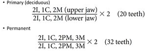
Dental Formula
The dental formula indicates the number and position of teeth in the upper and lower jaws for both primary and permanent dentition.
Dentition | Formula (per half jaw) | Total Teeth |
|---|---|---|
Primary (deciduous) | 2I, 1C, 2M (upper jaw) 2I, 1C, 2M (lower jaw) × 2 | 20 |
Permanent | 2I, 1C, 2PM, 3M (upper jaw) 2I, 1C, 2PM, 3M (lower jaw) × 2 | 32 |
Key: I = Incisor, C = Canine, PM = Premolar, M = Molar
Tooth Structure
Crown: Exposed part above gum; covered by enamel.
Root: Embedded in jawbone; attached by cement and periodontal ligament.
Enamel: Hardest substance in body; covers crown.
Dentin: Bonelike material under enamel.
Pulp cavity: Contains connective tissue, blood vessels, nerves.
Root canal: Extends through root; entry for vessels and nerves.
Pharynx and Esophagus
Food passes from mouth to pharynx (oropharynx, laryngopharynx) and then to esophagus. The esophagus is a muscular tube that propels food to the stomach.
Stomach
The stomach is a temporary storage tank that starts chemical breakdown of proteins. It converts food to chyme and delivers it to the small intestine. The mucosal barrier protects against harsh conditions.
Regions: Cardia, fundus, body, pyloric part.
Gastric glands: Mucous neck cells, parietal cells (HCl, intrinsic factor), chief cells (pepsinogen, lipase), enteroendocrine cells.
Small Intestine
The small intestine is the major organ of digestion and absorption. It is subdivided into duodenum, jejunum, and ileum, and has structural modifications (circular folds, villi, microvilli) to increase surface area.
Enterocytes: Absorptive cells with microvilli.
Goblet cells: Secrete mucus.
Paneth cells: Secrete antimicrobial agents.
Stem cells: Continuously divide to renew epithelium.
Large Intestine
The large intestine absorbs water, stores residues, and eliminates feces. It houses gut bacteria and is subdivided into cecum, appendix, colon, rectum, and anal canal.
Teniae coli: Longitudinal muscle bands.
Haustra: Pocketlike sacs.
Epiploic appendages: Fat-filled pouches.
Accessory Digestive Organs
Liver: Produces bile, processes nutrients, detoxifies blood.
Gallbladder: Stores and concentrates bile.
Pancreas: Produces digestive enzymes and bicarbonate.
Microscopic Anatomy of Liver and Pancreas
Liver lobules: Hexagonal units with plates of hepatocytes and central vein.
Portal triad: Branch of hepatic artery, portal vein, and bile duct.
Pancreatic acini: Clusters of cells secreting digestive enzymes.
Pancreatic Juices
Proteases: Digest proteins (trypsin, chymotrypsin, carboxypeptidase).
Amylase: Digests carbohydrates.
Lipases: Digests lipids.
Nucleases: Digests nucleic acids.
Additional info: The notes have been expanded with academic context and definitions to ensure completeness and clarity for college-level study.
