 Back
BackJoints and the Human Skeleton: Structure, Function, and Classification
Study Guide - Smart Notes
 Tailored notes based on your materials, expanded with key definitions, examples, and context.
Joints: Structure and Function
Introduction to Joints
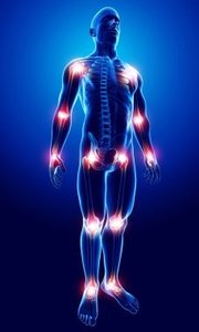
Joints, also known as articulations, are the locations where two or more bones meet. They are essential for movement, as bones themselves are rigid and cannot bend. The classification of joints is based on both their structural composition and their functional capacity for movement.
Structural Classification of Joints
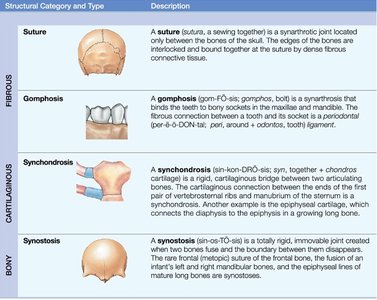
Joints are structurally categorized by the type of connective tissue that binds the bones and the presence or absence of a joint cavity. The four main structural types are:
Fibrous Joints: Bones are connected by fibrous connective tissue. Examples include sutures in the skull and gomphoses between teeth and jawbones.
Cartilaginous Joints: Bones are joined by cartilage. Examples include synchondroses (such as the epiphyseal plates in growing bones).
Bony Joints (Synostoses): Two bones fuse to form a single bone, as seen in the fusion of the frontal bone in the adult skull.
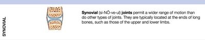
Synovial Joints: Bones are separated by a synovial cavity and connected by ligaments and a synovial membrane, allowing for a wide range of movement.
Functional Classification of Joints
Functionally, joints are classified by the degree of movement they allow:
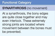
Synarthrosis: Immovable joints, such as sutures in the skull. These joints are extremely strong and prevent movement between bones.
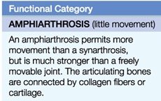
Amphiarthrosis: Slightly movable joints, such as the intervertebral discs. These joints are stronger than freely movable joints but allow limited movement.
Diarthrosis: Freely movable joints, such as the shoulder and hip. These joints are typically synovial and allow movement in one or more planes.
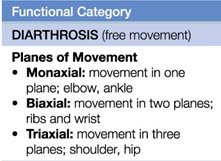
Planes of Movement in Diarthroses:
Monaxial: Movement in one plane (e.g., elbow, ankle)
Biaxial: Movement in two planes (e.g., ribs, wrist)
Triaxial: Movement in three planes (e.g., shoulder, hip)
The Axial Skeleton
Overview of the Axial Skeleton
The axial skeleton forms the central axis of the body and consists of the skull, vertebral column, and thoracic cage. It provides support and protection for the brain, spinal cord, and organs in the thoracic and abdominal cavities.
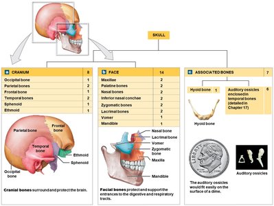
Skull and Associated Bones
The skull is composed of 22 bones: 8 cranial bones that protect the brain and 14 facial bones that form the structure of the face. Additionally, 7 other bones are associated with the skull: 6 auditory ossicles (involved in hearing) and 1 hyoid bone (supports the tongue).
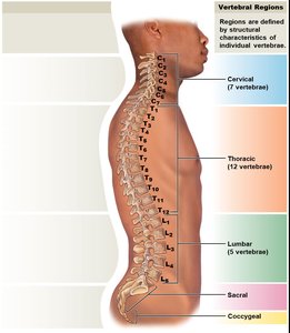
The Vertebral Column
The vertebral column (spine) connects the skull to the pelvis and is composed of 26 bones: 24 vertebrae, 1 sacrum, and 1 coccyx. It is divided into five regions:
Cervical (7 vertebrae)
Thoracic (12 vertebrae)
Lumbar (5 vertebrae)
Sacral (1 sacrum)
Coccygeal (1 coccyx)

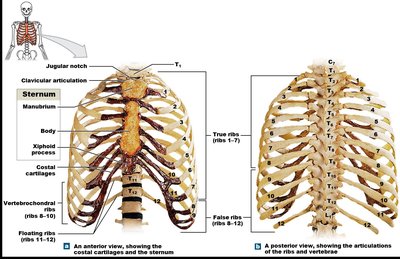
The Thoracic Cage
The thoracic cage consists of 24 ribs and the sternum. It protects vital organs in the thoracic cavity and provides attachment points for muscles involved in respiration and movement of the upper limbs. Ribs are classified as:
True ribs (1-7): Attach directly to the sternum via costal cartilage.
False ribs (8-12): Do not attach directly to the sternum. Ribs 8-10 are vertebrochondral, and ribs 11-12 are floating ribs.
The sternum is composed of three parts: the manubrium, body, and xiphoid process. Ossification and fusion of the sternum are not complete until at least age 25.
The Appendicular Skeleton
Overview of the Appendicular Skeleton
The appendicular skeleton includes the bones of the limbs and the girdles that attach them to the axial skeleton. It is responsible for movement and manipulation of the environment.
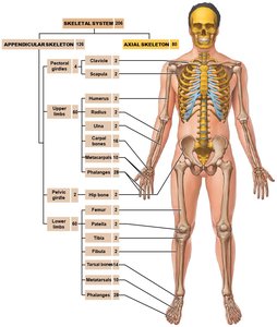
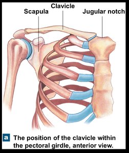
Pectoral Girdle
The pectoral girdle attaches the upper limbs to the axial skeleton. Each girdle consists of a clavicle and a scapula.
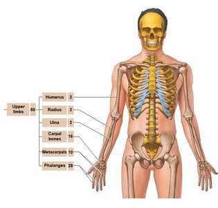
Upper Limbs
The upper limbs are composed of the humerus (arm), radius and ulna (forearm), carpals (wrist), metacarpals (hand), and phalanges (fingers).
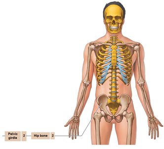
Pelvic Girdle
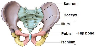
The pelvic girdle attaches the lower limbs to the axial skeleton and is formed by the paired hip bones (coxal bones), which consist of the ilium, ischium, and pubis.
Lower Limbs
The lower limbs include the femur (thigh), patella (kneecap), tibia and fibula (leg), tarsals (ankle), metatarsals (foot), and phalanges (toes).
Summary Table: Major Bones of the Human Skeleton
Region | Bones | Count |
|---|---|---|
Skull | Cranial, facial, auditory ossicles, hyoid | 29 |
Vertebral Column | Vertebrae, sacrum, coccyx | 26 |
Thoracic Cage | Sternum, ribs | 25 |
Pectoral Girdle | Clavicle, scapula | 4 |
Upper Limbs | Humerus, radius, ulna, carpals, metacarpals, phalanges | 60 |
Pelvic Girdle | Hip bones | 2 |
Lower Limbs | Femur, patella, tibia, fibula, tarsals, metatarsals, phalanges | 60 |
Key Take-Home Points
The skeletal system is composed of bones, joints, cartilages, and ligaments.
Bones are classified by shape and have unique markings for muscle and ligament attachment.
Bone is a connective tissue with a matrix of cells, collagen fibers, and calcium phosphate.
Bone growth and remodeling occur throughout life.
Joints are classified by structure (how bones are joined) and function (degree of movement).
The axial skeleton includes the skull, vertebral column, and thoracic cage.
The appendicular skeleton includes the pectoral and pelvic girdles and the bones of the limbs.
