Back
BackMajor Muscles of the Human Body: Origins, Insertions, Actions, and Nerves
Study Guide - Smart Notes
 Tailored notes based on your materials, expanded with key definitions, examples, and context.
Muscles of the Head and Face
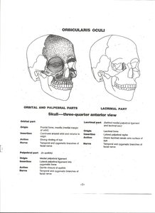
Orbicularis Oculi
The orbicularis oculi is a muscle responsible for closing the eyelids and facilitating blinking and squinting. It consists of orbital, palpebral, and lacrimal parts, each with distinct origins and insertions.
Origin: Frontal bone, maxilla, medial palpebral ligament, lacrimal bone
Insertion: Skin around orbit, lateral palpebral raphe, skin of eyelids
Action: Closes eyelids, compresses lacrimal sac
Nerve: Facial nerve (CN VII)
Orbicularis Oris
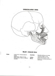
The orbicularis oris muscle encircles the mouth and is essential for movements such as puckering the lips, speaking, and eating.
Origin: Maxilla, mandible, and skin around the mouth
Insertion: Skin and mucous membrane of lips
Action: Closes and protrudes lips
Nerve: Facial nerve (CN VII)
Muscles of the Neck
Platysma
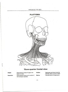
The platysma is a superficial muscle of the neck that assists in depressing the mandible and tensing the skin of the neck.
Origin: Subcutaneous fascia of upper chest and shoulder
Insertion: Lower border of mandible, skin and muscle at corner of mouth
Action: Depresses and draws lower lip laterally, tenses skin of neck
Nerve: Cervical branch of facial nerve
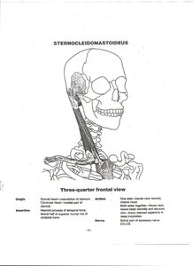
Sternocleidomastoideus
The sternocleidomastoid muscle is a prominent neck muscle involved in head rotation and flexion.
Origin: Sternum and clavicle
Insertion: Mastoid process of temporal bone, superior nuchal line of occipital bone
Action: Rotates head, flexes neck
Nerve: Accessory nerve (CN XI), cervical nerves
Muscles of the Chest and Shoulder
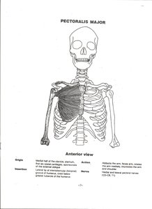
Pectoralis Major
The pectoralis major is a large chest muscle responsible for adducting and medially rotating the arm.
Origin: Medial half of clavicle, sternum, costal cartilages
Insertion: Lateral lip of intertubercular sulcus of humerus
Action: Adducts and medially rotates arm
Nerve: Medial and lateral pectoral nerves (C5–T1)
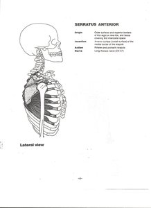
Serratus Anterior
The serratus anterior muscle stabilizes the scapula and assists in upward rotation.
Origin: Outer surfaces and superior borders of upper eight or nine ribs
Insertion: Anterior surface of medial border of scapula
Action: Protracts and rotates scapula
Nerve: Long thoracic nerve (C5–C7)
Muscles of the Abdomen
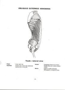
Obliquus Externus Abdominis
The external oblique muscle compresses the abdominal contents and aids in trunk rotation.
Origin: Lower eight ribs
Insertion: Iliac crest, abdominal aponeurosis
Action: Compresses abdomen, rotates trunk
Nerve: Lower six thoracic nerves
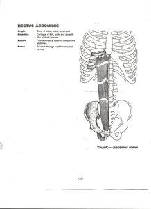
Rectus Abdominis
The rectus abdominis is a paired muscle running vertically on each side of the anterior wall of the human abdomen, important for flexing the lumbar spine.
Origin: Crest of pubis, pubic symphysis
Insertion: Costal cartilages of ribs 5–7, xiphoid process
Action: Flexes vertebral column, compresses abdomen
Nerve: Lower six thoracic nerves
Muscles of the Back
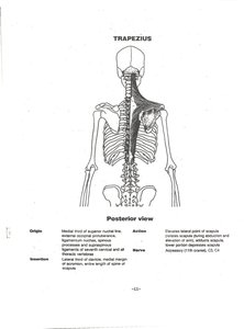
Trapezius
The trapezius muscle is a large, triangular muscle extending over the back of the neck and shoulders, responsible for moving, rotating, and stabilizing the scapula.
Origin: Occipital bone, ligamentum nuchae, spinous processes of C7–T12
Insertion: Lateral third of clavicle, acromion, spine of scapula
Action: Elevates, retracts, and rotates scapula
Nerve: Accessory nerve (CN XI), cervical nerves
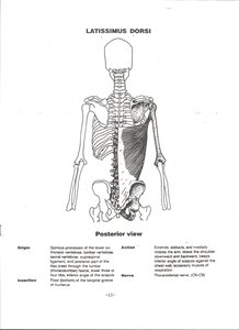
Latissimus Dorsi
The latissimus dorsi is a broad, flat muscle of the back that extends, adducts, and medially rotates the arm.
Origin: Spinous processes of T7–L5, iliac crest, lower ribs
Insertion: Intertubercular sulcus of humerus
Action: Extends, adducts, and medially rotates arm
Nerve: Thoracodorsal nerve (C6–C8)
Muscles of the Shoulder and Arm
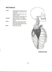
Deltoideus (Deltoid)
The deltoid muscle forms the rounded contour of the shoulder and is responsible for abduction, flexion, and extension of the arm.
Origin: Clavicle, acromion, spine of scapula
Insertion: Deltoid tuberosity of humerus
Action: Abducts, flexes, and extends arm
Nerve: Axillary nerve (C5, C6)
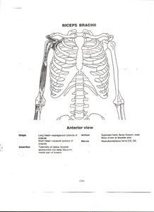
Biceps Brachii
The biceps brachii is a two-headed muscle of the upper arm, important for flexion and supination of the forearm.
Origin: Scapula (long head: supraglenoid tubercle; short head: coracoid process)
Insertion: Radial tuberosity, bicipital aponeurosis
Action: Flexes and supinates forearm
Nerve: Musculocutaneous nerve (C5, C6)
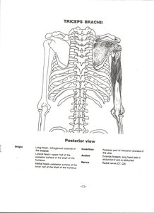
Triceps Brachii
The triceps brachii is a three-headed muscle of the upper arm, responsible for extension of the forearm at the elbow.
Origin: Scapula (long head), humerus (lateral and medial heads)
Insertion: Olecranon process of ulna
Action: Extends forearm
Nerve: Radial nerve (C7, C8)
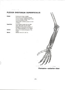
Flexor Digitorum Superficialis
The flexor digitorum superficialis is a muscle of the forearm that flexes the fingers.
Origin: Medial epicondyle of humerus, coronoid process of ulna, shaft of radius
Insertion: Middle phalanges of fingers 2–5
Action: Flexes middle phalanges of fingers
Nerve: Median nerve (C7, C8, T1)
Muscles of the Hip and Thigh
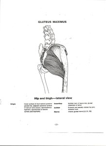
Gluteus Maximus
The gluteus maximus is the largest muscle of the buttocks, responsible for extension and lateral rotation of the hip.
Origin: Ilium, sacrum, coccyx
Insertion: Gluteal tuberosity of femur, iliotibial tract
Action: Extends and laterally rotates hip
Nerve: Inferior gluteal nerve (L5, S1, S2)
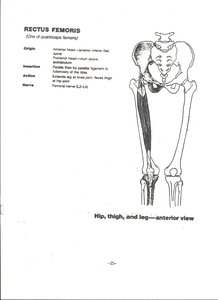
Rectus Femoris
The rectus femoris is one of the four quadriceps muscles, important for knee extension and hip flexion.
Origin: Anterior inferior iliac spine, ilium above acetabulum
Insertion: Patella, tibial tuberosity via patellar ligament
Action: Extends knee, flexes hip
Nerve: Femoral nerve (L2–L4)
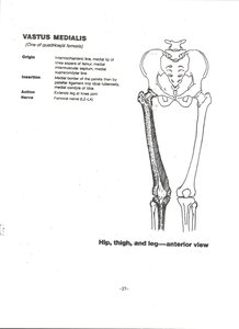
Vastus Medialis
The vastus medialis is another quadriceps muscle, crucial for knee extension.
Origin: Intertrochanteric line, medial lip of linea aspera
Insertion: Patella, tibial tuberosity via patellar ligament
Action: Extends knee
Nerve: Femoral nerve (L2–L4)
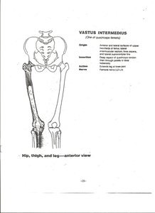
Vastus Intermedius
The vastus intermedius lies beneath the rectus femoris and also extends the knee.
Origin: Anterior and lateral surfaces of femur
Insertion: Patella, tibial tuberosity via patellar ligament
Action: Extends knee
Nerve: Femoral nerve (L2–L4)
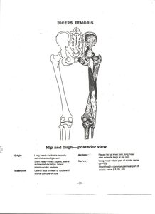
Biceps Femoris
The biceps femoris is a posterior thigh muscle, part of the hamstrings, responsible for knee flexion and hip extension.
Origin: Ischial tuberosity (long head), femur (short head)
Insertion: Head of fibula, lateral condyle of tibia
Action: Flexes knee, extends hip
Nerve: Sciatic nerve (L5, S1, S2)
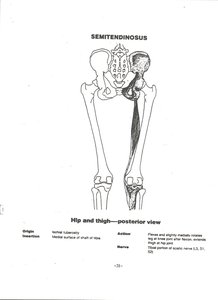
Semitendinosus
The semitendinosus is another hamstring muscle, aiding in knee flexion and hip extension.
Origin: Ischial tuberosity
Insertion: Medial surface of tibia
Action: Flexes knee, extends hip
Nerve: Sciatic nerve (L5, S1, S2)
Muscles of the Leg and Foot
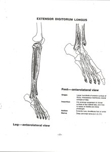
Extensor Digitorum Longus
The extensor digitorum longus is a muscle of the anterior leg that extends the toes and dorsiflexes the foot.
Origin: Lateral condyle of tibia, anterior surface of fibula
Insertion: Middle and distal phalanges of toes 2–5
Action: Extends toes, dorsiflexes foot
Nerve: Deep peroneal nerve (L4, L5, S1)
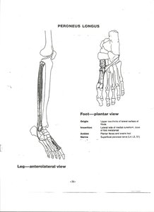
Peroneus Longus
The peroneus longus (fibularis longus) is a lateral leg muscle that everts and plantarflexes the foot.
Origin: Upper lateral shaft of fibula
Insertion: Base of first metatarsal, medial cuneiform
Action: Everts and plantarflexes foot
Nerve: Superficial peroneal nerve (L4, L5, S1)