 Back
BackMicroscopy and Staining Techniques in Microbiology
Study Guide - Smart Notes
 Tailored notes based on your materials, expanded with key definitions, examples, and context.
Observing Microorganisms Through a Microscope
Introduction to Microscopy
Microscopy is essential for studying microorganisms, which are too small to be seen with the naked eye. Various types of microscopes and staining techniques allow scientists to visualize and differentiate microbial structures and species.
Microscopy: The Instruments
Types of Microscopes
Simple Microscope: Contains a single lens, similar to a magnifying glass but with higher magnification.
Compound Light Microscope: Uses multiple lenses and visible light to magnify specimens.
Other Light Microscopy Types: Includes darkfield, phase-contrast, differential interference contrast (DIC), fluorescence, and confocal microscopy.
Parts of a Compound Light Microscope
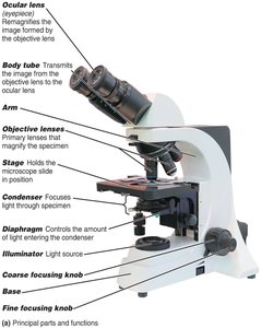
The compound light microscope consists of several key components that work together to magnify and resolve images of microorganisms.
Ocular lens (eyepiece): Remagnifies the image formed by the objective lens.
Objective lenses: Primary lenses that magnify the specimen.
Stage: Holds the microscope slide in position.
Condenser: Focuses light through the specimen.
Diaphragm: Controls the amount of light entering the condenser.
Illuminator: Light source.
Coarse and fine focusing knobs: Used to focus the image.
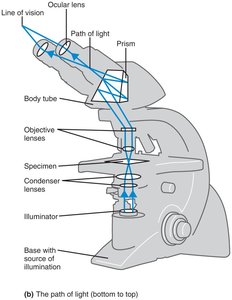
Path of Light in a Compound Microscope
Light passes from the illuminator through the condenser, specimen, objective lenses, and ocular lens, forming a magnified image for observation.
Total Magnification and Resolution
Total Magnification: Calculated as the product of the magnification of the objective lens and the ocular lens.
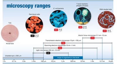
Resolution (Resolving Power): The ability to distinguish two points as separate entities. Higher resolution allows for clearer, more detailed images. The limit of resolution for a compound light microscope is about 0.2 μm, restricting its maximum useful magnification to approximately 1500x.
Wavelength: Shorter wavelengths of light provide greater resolution.
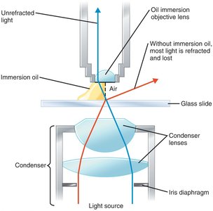
Refractive Index and Immersion Oil
The refractive index measures the light-bending ability of a medium. Immersion oil is used with high-power objective lenses to reduce light refraction and increase resolution.
Fluorescence Microscopy
Principles and Applications
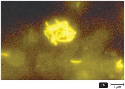
Fluorescence microscopy uses ultraviolet (UV) light to excite fluorescent dyes (fluorochromes) that emit visible light. This technique is valuable for detecting specific microorganisms, especially when using fluorescent-antibody (FA) techniques (immunofluorescence) for rapid and specific pathogen identification.
Fluorochromes: Dyes that fluoresce under UV light (e.g., Auramine O for Mycobacterium tuberculosis).
Immunofluorescence: Antibodies tagged with fluorochromes bind to specific microbes, causing them to fluoresce if present.
Electron Microscopy
Principles and Types
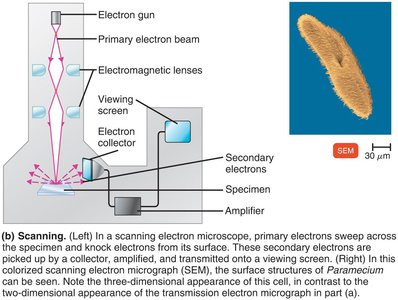
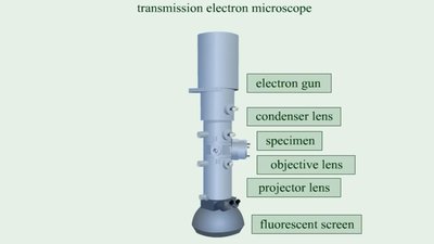
Electron microscopes use electron beams instead of light, providing much higher resolution due to the shorter wavelength of electrons. They are essential for visualizing viruses and internal cellular structures.
Transmission Electron Microscope (TEM): Electrons pass through ultrathin specimen sections, revealing internal structures. Magnification: 10,000–10,000,000x; resolution: 0.2 nm.
Scanning Electron Microscope (SEM): Electrons scan the specimen surface, producing detailed three-dimensional images. Magnification: 1,000–500,000x; resolution: 0.5 nm.
Preparation of Specimens for Light Microscopy
Staining and Fixation
Staining enhances contrast in microscopic images by coloring microorganisms. Before staining, a smear (thin film of specimen) is fixed to the slide by heat or chemicals to attach and preserve the cells.
Heat Fixation: Passing the slide through a flame.
Chemical Fixation: Using methanol to fix the smear.
Types of Dyes
Basic Dyes: Chromophore is a cation (e.g., crystal violet, methylene blue, safranin); stains bacterial cells directly.
Acidic Dyes: Chromophore is an anion (e.g., eosin, acid fuchsin, nigrosin); stains the background (negative staining).
Simple Stains
Simple stains use a single basic dye to highlight the entire microorganism, making cell shapes and structures visible. A mordant may be used to intensify the stain.
Differential Stains
Gram Stain
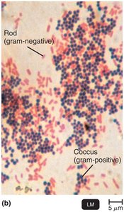
The Gram stain differentiates bacteria into gram-positive and gram-negative groups based on cell wall structure. It is a critical technique in medical microbiology for bacterial identification and treatment guidance.
Gram-Positive: Thick peptidoglycan cell wall; stains purple.
Gram-Negative: Thin peptidoglycan wall and outer membrane; stains pink/red.
Step | Gram-Positive | Gram-Negative |
|---|---|---|
Primary Stain: Crystal Violet | Purple | Purple |
Mordant: Gram’s Iodine | Purple | Purple |
Decolorizing Agent: Alcohol/Acetone | Purple | Colorless |
Counterstain: Safranin | Purple | Pink/Red |

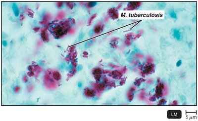
Acid-Fast Stain
The acid-fast stain identifies bacteria with waxy cell walls (e.g., Mycobacterium, Nocardia). Acid-fast bacteria retain the primary stain (carbolfuchsin) even after decolorization with acid-alcohol.
Step | Acid-Fast | Non–Acid-Fast |
|---|---|---|
Primary Stain: Carbolfuchsin | Red | Red |
Decolorizing Agent: Acid-Alcohol | Red | Colorless |
Counterstain: Methylene Blue | Red | Blue |
Special Stains
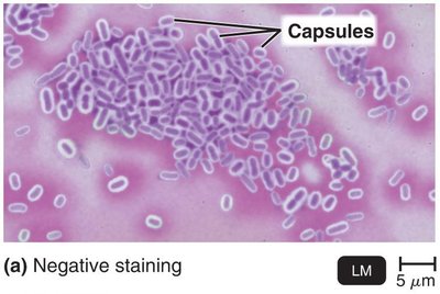
Capsule Stain (Negative Staining)
Capsule stains highlight the gelatinous outer covering of some bacteria, which do not accept most dyes. Negative staining with India ink or nigrosin contrasts the background, while a simple stain colors the cell, leaving the capsule as a clear halo.
Endospore Staining
Endospores are resistant, dormant structures that require special staining. The Schaeffer-Fulton method uses malachite green (with heat) as the primary stain and safranin as the counterstain. Endospores appear green within red or pink cells.

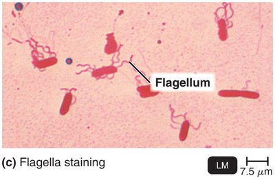
Flagella Staining
Flagella are thin structures of locomotion that are not visible with standard light microscopy. Special stains with mordant and carbolfuchsin thicken the flagella, making them visible and allowing determination of their number and arrangement.
Additional info: These microscopy and staining techniques are foundational for identifying and studying microorganisms in clinical and research settings. Mastery of these methods is essential for microbiology, pathology, and related biomedical sciences.
