 Back
BackMini-Textbook Study Notes: Skeletal System, Muscular System, and Nervous System
Study Guide - Smart Notes
 Tailored notes based on your materials, expanded with key definitions, examples, and context.
Bones and Skeletal Tissues
Bone Development and Ossification
Bone development, also known as osteogenesis or ossification, is the process by which new bone is formed. This process begins in the second month of embryonic development and continues throughout life, including postnatal growth and bone remodeling. The process is regulated by hormones such as pituitary growth hormone (GH), testosterone, and estrogen. Disorders such as giantism and dwarfism result from imbalances in these regulatory mechanisms.
Osteogenesis: Formation of bone tissue.
Postnatal bone growth: Occurs until early adulthood.
Bone remodeling and repair: Lifelong process.
Types of Ossification
There are two primary types of ossification:
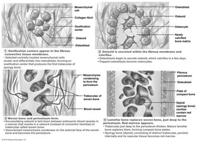
Intramembranous Ossification: Bone develops from a fibrous membrane, forming flat bones such as the clavicles and cranial bones.
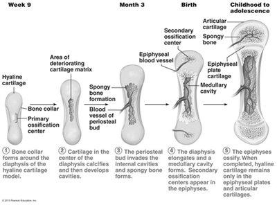
Endochondral Ossification: Bone forms by replacing hyaline cartilage, creating most of the skeleton except the skull and clavicle.
Intramembranous Ossification Steps
Ossification centers appear in the fibrous connective tissue.
Osteoblasts secrete bone matrix.
Woven bone and periosteum form.
Bone collar of compact bone and red marrow appear.
Endochondral Ossification Steps
Bone collar forms around hyaline cartilage model.
Cavitation of hyaline cartilage occurs.
Periosteal bud invades internal cavities, forming spongy bone.
Medullary cavity forms; secondary ossification centers appear in epiphyses.
Epiphyses ossify, leaving epiphyseal plates and articular cartilages.
Bone Growth and Remodeling
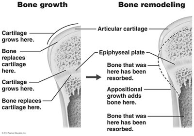
Bone growth occurs through interstitial (length) and appositional (width) mechanisms. Remodeling is a dynamic process involving osteoblasts (bone formation) and osteoclasts (bone resorption).
Interstitial growth: Increase in length of long bones.
Appositional growth: Increase in thickness; remodeling by osteoblasts and osteoclasts.
Bone Homeostasis: Remodeling and Repair
Bone is constantly remodeled and repaired. About 5-7% of bone is recycled weekly. Spongy bone is replaced every 3-4 years, and compact bone every 10 years.
Bone deposit: Requires protein, vitamins C, D, A, calcium, phosphorus, magnesium, and manganese.
Bone resorption: Osteoclasts digest organic matrix and convert calcium salts into soluble forms.
Hormonal Control of Blood Calcium
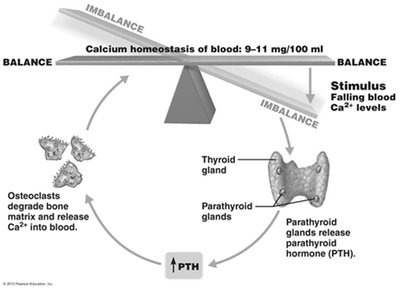
Calcium is essential for nerve transmission, muscle contraction, blood coagulation, gland secretion, and cell division. Blood calcium levels are regulated by parathyroid hormone (PTH) and calcitonin.
PTH: Increases blood calcium by stimulating osteoclasts.
Calcitonin: Lowers blood calcium by inhibiting osteoclasts.
Fracture Repair
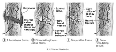
Bone fractures are repaired through a four-step process:
Hematoma forms.
Fibrocartilaginous callus forms.
Bony callus forms.
Bone remodeling occurs.
Muscular System and Muscle Tissue
Sarcomere Anatomy
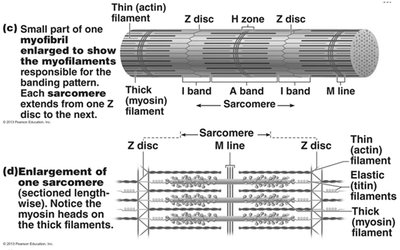
The sarcomere is the smallest contractile unit of a muscle fiber, defined as the region between two Z discs. It is composed of thick (myosin) and thin (actin) myofilaments.
Thick filaments: Run the length of the A band.
Thin filaments: Run the length of the I band and partway into the A band.
Z disc: Anchors thin filaments and connects myofibrils.
H zone: Lighter midregion where filaments do not overlap.
M line: Holds adjacent thick filaments together.
Muscle Contraction: Sliding Filament Model
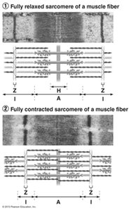
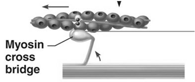
Muscle contraction occurs when myosin heads bind to actin, detach, and bind again, propelling thin filaments toward the M line. This shortens the sarcomere and the muscle fiber.
Neuromuscular Junction and Action Potential
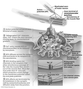
The neuromuscular junction is where a motor neuron stimulates a muscle fiber. Acetylcholine (ACh) is released, binds to receptors, and generates an action potential in the sarcolemma.
Activation: Neural stimulation at the neuromuscular junction.
Excitation-contraction coupling: Action potential propagation and Ca2+ release.
Destruction of Acetylcholine
ACh effects are terminated by acetylcholinesterase, preventing continued muscle contraction without further stimulation.
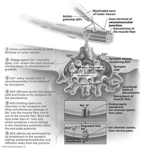
Generation of Action Potential
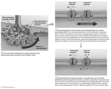
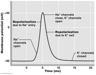
Action potentials are generated by the opening of voltage-gated Na+ channels, leading to depolarization, followed by repolarization as K+ channels open.
Role of Calcium in Contraction
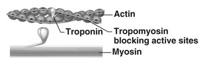
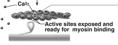
Calcium ions are essential for muscle contraction. At low Ca2+ concentrations, tropomyosin blocks actin's active sites. At higher concentrations, Ca2+ binds to troponin, moving tropomyosin and allowing myosin to bind to actin.
Cross Bridge Cycle
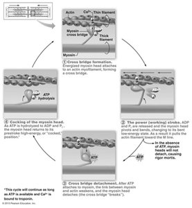
The cross bridge cycle continues as long as Ca2+ and ATP are present. It involves cross bridge formation, power stroke, detachment, and cocking of the myosin head.
Muscle Mechanics and Motor Units
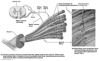
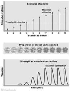
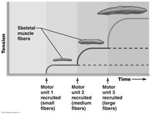
Muscle contraction produces tension. Motor units consist of a motor neuron and all the muscle fibers it innervates. Different motor units contract asynchronously to prevent fatigue.
Muscle Twitch and Graded Responses
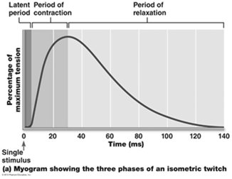
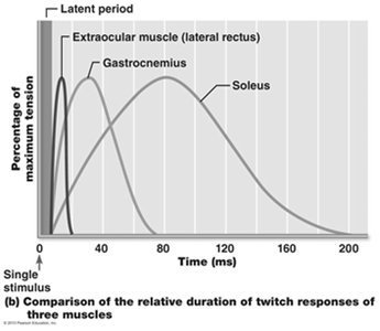
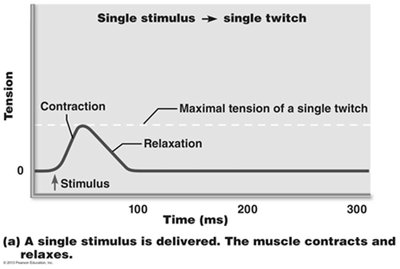
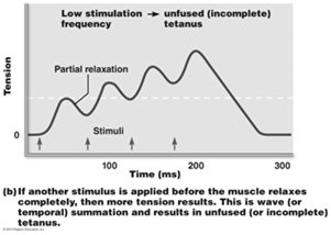
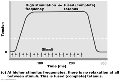
A muscle twitch is the response to a single, brief stimulus. It has three phases: latent period, contraction, and relaxation. Graded responses are achieved by changing the frequency and strength of stimulation.
Muscle Tone and Types of Contraction
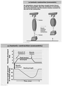
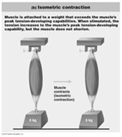
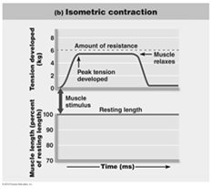
Muscle tone is a constant, slightly contracted state. Isotonic contractions involve muscle shortening (concentric) or lengthening (eccentric). Isometric contractions increase tension without changing muscle length.
Muscle Metabolism: Energy for Contraction
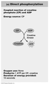
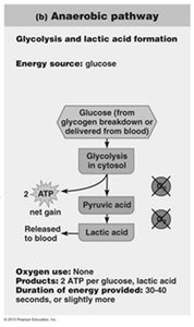
ATP is the direct energy source for muscle contraction. It is regenerated by direct phosphorylation, anaerobic glycolysis, and aerobic respiration.
Nervous System Fundamentals
Neuron Function and Principles of Electricity
Neurons generate action potentials in response to stimuli. Key electrical concepts include voltage, current, and resistance.
Voltage (V): Potential energy from separated charges.
Current (I): Flow of electrical charge.
Resistance (R): Hindrance to charge flow.
Membrane Ion Channels
Membrane proteins serve as ion channels, which can be leakage (always open) or gated (chemically, voltage, or mechanically gated).
Resting Membrane Potential
The resting membrane potential is typically -70 mV in neurons, maintained by differences in ionic composition and permeability, and stabilized by the sodium-potassium pump.
Changes in Membrane Potential
Membrane potential changes act as signals. Depolarization decreases membrane potential, while hyperpolarization increases it.
Action Potential Generation
Action potentials are brief reversals of membrane potential, involving depolarization, repolarization, and hyperpolarization. They are generated by voltage-gated channels and do not decrease in magnitude over distance.
Threshold and Propagation
Action potentials are all-or-none events, triggered when membrane potential reaches threshold. Propagation occurs as Na+ influx depolarizes adjacent areas, ensuring one-way transmission.
Stimulus Intensity Coding and Refractory Periods
The CNS codes stimulus intensity by the frequency of action potentials. Absolute and relative refractory periods ensure unidirectional transmission and limit firing rate.
Conduction Velocity and Myelination
Conduction velocity depends on axon diameter and myelination. Myelinated axons conduct impulses faster via saltatory conduction.
Synapses and Neurotransmitters
Synapses are junctions for information transfer. Chemical synapses release neurotransmitters, which bind to postsynaptic receptors and generate graded potentials. Neurotransmitter effects are terminated by degradation, reuptake, or diffusion.
Peripheral Nervous System
Sensory Receptors and Integration
Sensory receptors respond to environmental changes, triggering graded potentials and nerve impulses. Receptors are classified by stimulus type, location, and structural complexity.
Mechanoreceptors: Touch, pressure, vibration, stretch.
Thermoreceptors: Temperature changes.
Photoreceptors: Light energy.
Chemoreceptors: Chemicals.
Nociceptors: Pain-causing stimuli.
Classification by Location
Exteroceptors: Respond to external stimuli.
Interoceptors: Respond to internal stimuli.
Proprioceptors: Respond to stretch in muscles and joints.
Structural Complexity
Complex receptors: Special sense organs.
Simple receptors: General senses; unencapsulated or encapsulated dendritic endings.
Reflexes and Reflex Arc
Reflexes are rapid, involuntary responses to stimuli. The reflex arc consists of receptor, sensory neuron, integration center, motor neuron, and effector.
Autonomic Nervous System (ANS)
Divisions and Functions
The ANS consists of motor neurons that innervate smooth and cardiac muscle and glands. It operates subconsciously and is divided into sympathetic (fight or flight) and parasympathetic (rest and digest) divisions.
Efferent Pathways and Neurotransmitters
Somatic nervous system: Single, heavily myelinated motor neuron; releases ACh.
ANS: Two-neuron chain; preganglionic (myelinated) and postganglionic (unmyelinated); releases ACh or NE.
Neurotransmitter Receptors
Cholinergic receptors: Bind ACh (nicotinic and muscarinic).
Adrenergic receptors: Bind NE and epinephrine (alpha and beta subclasses).
Effects of Drugs
Atropine: Anticholinergic; blocks muscarinic receptors.
Neostigmine: Inhibits acetylcholinesterase; treats myasthenia gravis.
Beta-blockers: Attach to beta receptors; used in asthma and other conditions.
Additional info: Academic context was added to clarify mechanisms, definitions, and examples for each topic. Tables referenced in the original material were not included due to lack of direct content, but key comparisons and classifications were described in text.
