 Back
BackMotor Control: Functional Anatomy and Pathways of Voluntary Movement
Study Guide - Smart Notes
 Tailored notes based on your materials, expanded with key definitions, examples, and context.
Motor Control: Functional Anatomy and Pathways of Voluntary Movement
Introduction to Motor Control
The nervous system orchestrates voluntary body movements through a complex network of brain regions, spinal cord tracts, and feedback mechanisms. Understanding motor control involves identifying the key anatomical structures, their roles, and the pathways that transmit motor commands from the brain to skeletal muscles.
Functional Organization of the Nervous System
Autonomic vs. Somatic Nervous System
Autonomic Nervous System (ANS): Controls involuntary functions (e.g., heart rate, digestion) and maintains homeostasis by regulating smooth muscle, cardiac muscle, and glands.
Somatic Nervous System (SNS): Governs voluntary motor responses via skeletal muscles. While primarily under conscious control, some actions (e.g., reflexes, learned motor skills) can become automatic or unconscious.
Key Functions of the Nervous System: Sensation, integration, and response. Control can be further classified as autonomic (involuntary) or somatic (voluntary).
Functional Anatomy of Motor Control
Cerebral Cortex and Motor Areas
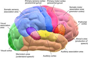
The frontal lobe is central to motor control, with distinct regions responsible for planning, initiating, and executing movements.
Prefrontal Cortex: Executive functions, goal-directed behavior, attention, and working memory.
Premotor Cortex: Controls core muscles for posture during movement; lateral location.
Supplemental Motor Area: Medial and superior; plans and coordinates learned, sequential movements.
Frontal Eye Fields: Controls voluntary eye movements in response to visual stimuli.
Broca’s Area: Controls muscles involved in speech production.
Primary Motor Cortex: Located in the precentral gyrus; initiates voluntary muscle contractions and is organized as a motor homunculus, with more cortical space devoted to muscles requiring fine control (e.g., fingers, face).
Descending Motor Pathways
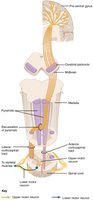
Motor commands travel from the cortex to the spinal cord via descending tracts, primarily the corticospinal and corticobulbar tracts.
Upper Motor Neurons: Originate in the primary motor cortex; their axons form descending tracts.
Corticobulbar Tract: Projects to cranial motor nuclei in the brainstem; controls muscles of the face, head, and neck (mostly ipsilateral projections).
Corticospinal Tract: Projects to the ventral horn of the spinal cord; controls muscles of the torso and limbs (mostly contralateral projections due to decussation at the pyramidal decussation in the medulla).
Lateral Corticospinal Tract: Controls appendicular muscles (arms and legs); fibers cross at the pyramidal decussation.
Anterior Corticospinal Tract: Controls axial muscles (trunk); fibers may be bilateral, with some decussating at the spinal cord level.
Extrapyramidal System
Extrapyramidal tracts originate outside the pyramidal (corticospinal) system and are influenced by subcortical structures:
Tectospinal Tract: Mediates postural movements in response to visual stimuli (from the superior colliculus/tectum).
Reticulospinal Tract: Influences posture, locomotion, muscle tone, and some autonomic functions.
Vestibulospinal Tract: Modulates posture and balance based on equilibrium information from the vestibular system.
Rubrospinal Tract: Provides cerebellar corrections to motor commands.
Role of the Cerebellum and Basal Nuclei
Cerebellum: Compares intended movement (from cortex) with proprioceptive feedback; corrects errors via the rubrospinal tract. Essential for coordination and balance.
Basal Nuclei: Involved in motor planning and modulation of movement initiation.
Ventral Horn of the Spinal Cord
Lower motor neurons (somatic motor neurons) reside in the ventral horn and directly innervate skeletal muscles. Their axons exit via the ventral root and can be very long, especially for distal muscles.
Phases of Voluntary Movement
Overview of Voluntary Movement Phases
Voluntary movements are complex and involve multiple anatomical regions. They can be divided into three main phases:
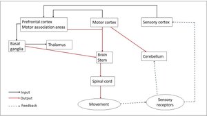
Planning: Prefrontal cortex integrates sensory information and plans movement with input from the cerebellum and basal nuclei.
Initiation: Premotor cortex and supplemental motor area coordinate and initiate the movement plan.
Execution: Primary motor cortex sends commands down the corticospinal tract to lower motor neurons, which activate skeletal muscles. The cerebellum and basal nuclei provide ongoing feedback for posture and balance.
Example: Throwing a Baseball
Planning: The pitcher’s prefrontal cortex assesses the environment and decides on the pitch type, integrating sensory input from the cortex, cerebellum, basal nuclei, and spinal cord.
Initiation: Premotor and supplemental motor areas, basal ganglia, and thalamus initiate the pitch by activating upper motor neurons in the primary motor cortex.
Execution: Upper motor neurons stimulate lower motor neurons, which contract specific muscles (e.g., infraspinatus, teres minor, supraspinatus, subscapularis) via the appropriate nerves to perform the throwing motion.
Summary Table: Roles of Brain Structures in Motor Control
Nervous System Structure | Role in Motor Control |
|---|---|
Brain stem | Maintains posture, integrates reflexes, and coordinates basic movement patterns |
Thalamus | Relays and modifies motor signals between the cerebellum, basal nuclei, and cortex |
Motor cortex | Initiates and executes voluntary movements |
Cerebellum | Coordinates movement, corrects errors, and maintains balance and posture |
Key Definitions and Concepts
Upper Motor Neuron: Neuron originating in the cortex or brainstem, projecting to lower motor neurons.
Lower Motor Neuron: Neuron in the spinal cord or brainstem that directly innervates skeletal muscle.
Decussation: Crossing of nerve fibers from one side of the CNS to the other (e.g., pyramidal decussation in the medulla).
Homunculus: Topographical map of the body on the motor cortex, reflecting the degree of motor control.
Summary of Motor Control Pathways
Tract | Origin | Decussation | Target Muscles | Function |
|---|---|---|---|---|
Corticobulbar | Motor cortex | Mostly ipsilateral | Face, head, neck | Facial and cranial muscle control |
Lateral corticospinal | Motor cortex | Pyramidal decussation (medulla) | Arms, legs | Fine voluntary movement of limbs |
Anterior corticospinal | Motor cortex | Some fibers decussate at spinal cord | Trunk | Posture and gross movements |
Tectospinal | Midbrain (tectum) | Varies | Neck, trunk | Postural responses to visual stimuli |
Reticulospinal | Reticular formation | Varies | Trunk, proximal limbs | Posture, locomotion, muscle tone |
Vestibulospinal | Vestibular nuclei | Varies | Trunk, limbs | Balance and equilibrium |
Summary
Motor control involves a hierarchy of brain regions and spinal pathways that plan, initiate, and execute voluntary movements.
Descending tracts transmit motor commands, while feedback from the cerebellum and sensory systems ensures coordination and accuracy.
Understanding these pathways is essential for diagnosing and treating motor disorders.
