 Back
BackMuscle Metabolism, Fatigue, and Fiber Types: Study Notes for ANP College Students
Study Guide - Smart Notes
 Tailored notes based on your materials, expanded with key definitions, examples, and context.
Muscle Metabolism and Energy for Contraction
ATP Production in Muscle Fibers
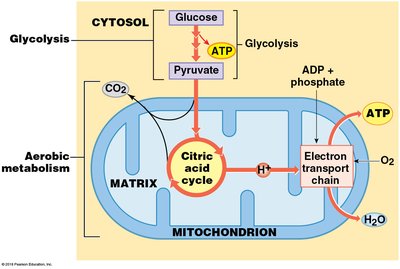
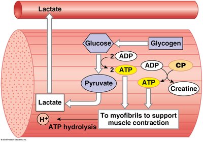
Muscle contraction requires significant amounts of ATP, which can be generated through both anaerobic and aerobic pathways. The efficiency and duration of muscle activity depend on the metabolic processes used to produce ATP.
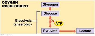
Glycolysis (Anaerobic Metabolism): Occurs in the cytosol without oxygen, breaking down glucose into pyruvate and producing 2 ATP molecules per glucose.
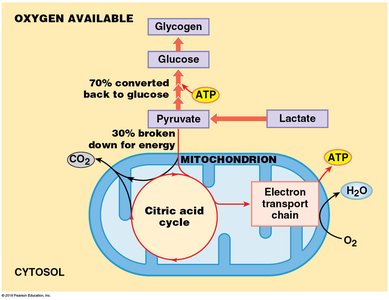
Aerobic Metabolism: Takes place in mitochondria, using oxygen to produce up to 15 ATP per pyruvate, primarily through the citric acid cycle and electron transport chain.
Energy Reserves in Skeletal Muscle Fibers
Muscle fibers store energy in several forms to support contraction:
Glycogen: The primary energy reserve, making up to 1.5% of muscle weight.
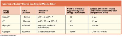
Free ATP: Present in minimal amounts, sufficient for only a few muscle twitches.
Creatine Phosphate (CP): Provides energy for short bursts (about 15 seconds) by donating phosphate to ADP to form ATP.
Energy Source | Initial Quantity | Utilization Process | Number of Twitches Supported | Duration of Contraction Supported |
|---|---|---|---|---|
Free ATP | 3 mmol | ATP → ADP + Pi | 10 | 2 sec |
CP | 20 mmol | ADP + CP → ATP + C | 70 | 15 sec |
Glycogen (anaerobic) | 100 mmol | Glycolysis | 670 | 130 sec |
Glycogen (aerobic) | 100 mmol | Aerobic metabolism | 12,000 | 2400 sec (40 min) |
Muscle Metabolism at Different Activity Levels
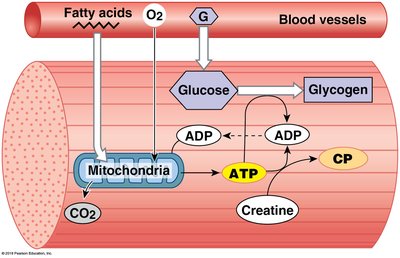
At Rest: Low ATP demand; mitochondria produce surplus ATP, which is used to build up reserves of creatine phosphate and glycogen. Fatty acids and glucose are absorbed from the bloodstream.
Moderate Activity: ATP demand increases; aerobic metabolism of pyruvate (from glycolysis) is the main source of ATP. Oxygen consumption rises, and fatigue does not occur until energy reserves are depleted.
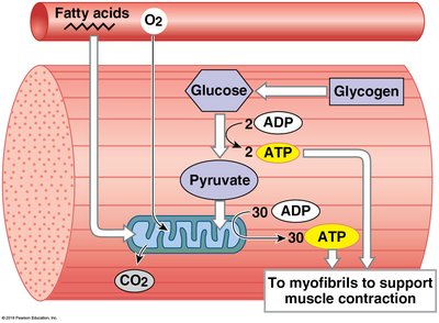
Peak Activity: ATP demand is very high; mitochondria provide about one-third of ATP, while the rest comes from anaerobic glycolysis. Excess pyruvate is converted to lactate, leading to lactic acidosis and muscle fatigue.
Muscle Fatigue and Recovery
Causes and Effects of Muscle Fatigue
Muscle fatigue occurs when a muscle can no longer perform at the required level. A major factor is decreased pH due to lactic acid accumulation, which impairs calcium binding and enzyme activity.
Glycolysis during Oxygen Insufficiency: When oxygen is limited, glycolysis is the only source of ATP. It is less efficient and leads to lactic acid buildup, lowering pH and causing fatigue.
Recovery Period and the Cori Cycle
During recovery, oxygen becomes available, and lactate is converted back to pyruvate. Pyruvate can be used to generate ATP or be recycled to glucose/glycogen. Most ATP is produced aerobically, which is more efficient than glycolysis.
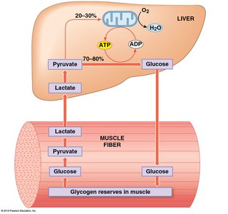
Cori Cycle: Lactate produced in muscles is transported to the liver, converted to pyruvate, and then to glucose, which returns to muscles to replenish glycogen stores.
Oxygen Debt (EPOC)
Oxygen debt, or excess post-exercise oxygen consumption (EPOC), is the amount of oxygen required to restore muscles to pre-exertion conditions. This includes replenishing ATP, creatine phosphate, and glycogen, as well as converting excess lactate to glucose in the liver.
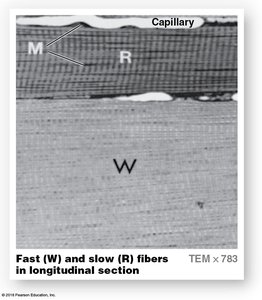
Skeletal Muscle Fiber Types
Classification of Muscle Fibers
Skeletal muscle fibers are classified based on their contraction speed, metabolic pathways, and resistance to fatigue:
Fast Fibers: Large diameter, few mitochondria, low myoglobin, rapid and powerful contractions, fatigue quickly (anaerobic metabolism).
Slow Fibers: Small diameter, many mitochondria, high myoglobin, slow but sustained contractions, fatigue-resistant (aerobic metabolism).
Intermediate Fibers: Characteristics between fast and slow fibers; more fatigue-resistant than fast fibers, but less than slow fibers.
Property | Fast Fibers | Slow Fibers | Intermediate Fibers |
|---|---|---|---|
Diameter | Large | Small | Intermediate |
Color | White | Red | Pink |
Myoglobin | Low | High | Low |
Capillaries | Few | Dense | Intermediate |
Mitochondria | Few | Many | Intermediate |
Fatigue Resistance | Low | High | Intermediate |
Muscle Fiber Distribution and Adaptation
Most muscles contain a mix of fiber types, reflecting their functional demands. The proportion of fast and slow fibers is genetically determined, but training can alter the ratio of intermediate fibers.
Muscle Hypertrophy, Atrophy, and Paralysis
Muscle Hypertrophy
Hypertrophy is the enlargement of muscle due to repeated, exhaustive stimulation. It results from increased mitochondria, glycogen, myofibrils, and myofilaments, leading to greater strength.
Muscle Atrophy
Atrophy is the reduction in muscle size, tone, and power due to decreased stimulation, aging, paralysis, or immobilization. Initially reversible, prolonged atrophy can lead to irreversible muscle fiber loss.

Muscle Paralysis and Disease
Muscular Dystrophy: Inherited diseases causing muscle weakness and deterioration (e.g., Duchenne and Becker muscular dystrophy).
Polio: Viral infection attacking CNS motor neurons, leading to atrophy and paralysis.
Tetanus: Bacterial toxin suppresses inhibition of motor neurons, causing sustained contractions.
Botulism: Bacterial toxin blocks acetylcholine release, resulting in paralysis.
Myasthenia Gravis: Autoimmune disease causing loss of acetylcholine receptors, leading to progressive weakness.
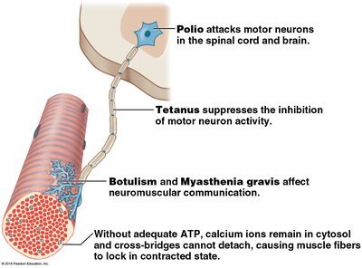
Rigor Mortis
Rigor mortis is the generalized muscle contraction after death, caused by ATP depletion and sustained calcium presence in the sarcoplasm. It ends as muscle tissue decomposes.
Additional info: The above notes integrate and expand upon the provided material, ensuring a comprehensive, exam-ready summary for ANP college students.
