 Back
BackMuscle Tissue and Physiology: Structure, Function, and Mechanisms
Study Guide - Smart Notes
 Tailored notes based on your materials, expanded with key definitions, examples, and context.
Muscle Tissue and Physiology
Overview of Muscle Tissue
Muscle tissue is specialized for contraction and is essential for movement, posture, joint stabilization, heat generation, and regulation of material flow through hollow organs. Muscle cells, or myocytes, are surrounded by an extracellular matrix called the endomysium, which helps transmit tension and maintain structural integrity.
Muscle Tension: The force generated by muscle tissue, responsible for movement and other physiological functions.
Types of Muscle Tissue: Skeletal, Cardiac, and Smooth muscle, each with distinct structures and functions.
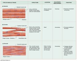
Types of Muscle Tissue
Skeletal Muscle Tissue
Skeletal muscle fibers are long, cylindrical, striated, and multinucleated. They are arranged parallel to each other and are mostly attached to the skeleton. Skeletal muscle is under voluntary control and must be stimulated by the nervous system.
Structure: Long, thin, striated, multinucleated cells.
Location: Attached to bones.
Function: Produces movement of the body.
Cardiac Muscle Tissue
Cardiac muscle cells are shorter, branched, and usually have a single nucleus. They are striated and connected by intercalated discs, which contain gap and tight junctions, allowing the heart to contract as a unit. Cardiac muscle is involuntary and found only in the heart.
Structure: Short, branched, striated cells with intercalated discs.
Location: Heart.
Function: Pumps blood throughout the body.
Smooth Muscle Tissue
Smooth muscle cells are long, flattened, and have two pointed ends with a single, centrally located nucleus. They are found lining hollow organs, in the eyes, skin, and ducts of certain glands. Smooth muscle is involuntary and many cells are linked by gap junctions.
Structure: Non-striated, spindle-shaped cells.
Location: Walls of hollow organs, eyes, skin, glands.
Function: Changes diameter of tubes, moves materials through organs.
Properties of Muscle Cells
Muscle cells possess unique properties that enable their function:
Contractility: Ability to contract and generate force.
Excitability (Responsivity): Ability to respond to stimuli (chemical, mechanical, or electrical).
Conductivity: Ability to conduct electrical charges across the plasma membrane.
Distensibility: Ability to be stretched without rupturing.
Elasticity: Ability to return to original length after being stretched.
Structure of Muscle Cells
Muscle cells share many organelles with other cells but have specialized structures:
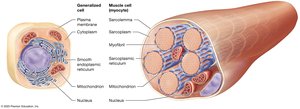
Sarcoplasm: Cytoplasm of a muscle cell.
Sarcolemma: Plasma membrane of a muscle cell.
Myofibrils: Bundles of specialized proteins responsible for contraction, making up 50–80% of cell volume.
Sarcoplasmic Reticulum (SR): Modified smooth endoplasmic reticulum that stores and releases calcium ions.
Structure of the Skeletal Muscle Fiber
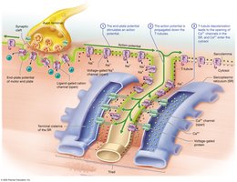
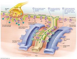
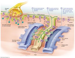
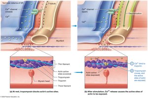
Skeletal muscle fibers are long, cylindrical, multinucleated, and striated. They arise from the fusion of multiple embryonic myoblasts. The sarcolemma forms inward extensions called transverse tubules (T-tubules) that surround each myofibril, forming a tunnel-like network filled with extracellular fluid. Flanking each T-tubule are enlarged portions of the SR called terminal cisternae; together, a T-tubule and two terminal cisternae form a triad.
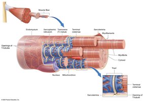
Structure of the Myofibril
Myofibrils are composed of three types of myofilaments:
Thick Filaments: Composed of myosin, with two globular heads and a tail. The heads bind to actin during contraction.
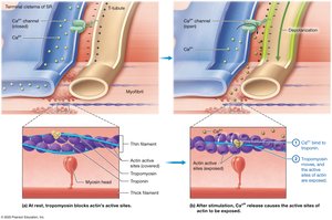
Thin Filaments: Composed of actin (with active sites for myosin binding), tropomyosin (covers active sites at rest), and troponin (regulates tropomyosin position).
Elastic Filaments: Composed of titin, which provides elasticity and helps the muscle return to its original length.
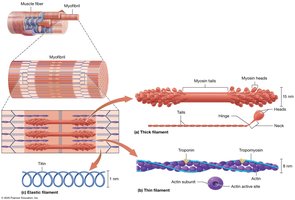
Myofilament Arrangement and the Sarcomere
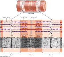
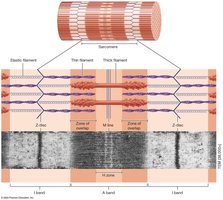
The arrangement of myofilaments creates the striated appearance of skeletal and cardiac muscle. The sarcomere is the functional unit of contraction, defined by the area between two Z-discs.
I band: Light region containing only thin filaments.
A band: Dark region containing thick filaments (and some thin filaments).
H zone: Middle of the A band, only thick filaments.
M line: Structural proteins holding thick filaments in place.
Z-discs: Anchor thin filaments and elastic filaments; attach myofibrils to one another.
The Sliding-Filament Mechanism of Contraction
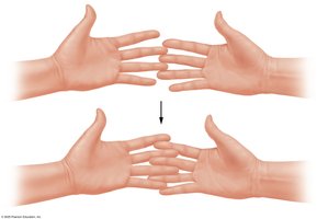
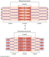
Muscle contraction occurs via the sliding-filament mechanism, where thin filaments slide past thick filaments, shortening the sarcomere and generating tension. The I bands and H zone narrow, while the A band remains unchanged.
Myosin heads pull thin filaments toward the M line, bringing Z-discs closer together.
None of the filaments themselves shorten; only their relative positions change.
Membrane Potential in Muscle Cells
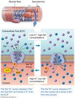
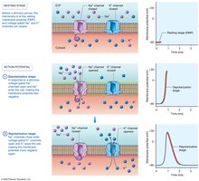
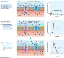
Muscle fibers maintain a resting membrane potential due to the distribution of ions across the sarcolemma. The sodium-potassium pump (Na+/K+ ATPase) maintains this gradient by moving three sodium ions out and two potassium ions into the cell, requiring ATP.
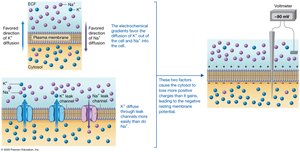
Resting membrane potential: About −90 mV in muscle fibers.
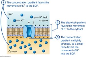
Ion channels: Leak channels (always open) and gated channels (open in response to stimuli).
Electrochemical gradient: Determines ion movement across the membrane.
Action Potentials
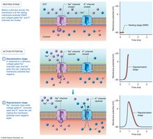
An action potential is a rapid, temporary change in membrane potential, essential for muscle contraction. It involves two main phases:
Depolarization: Voltage-gated sodium channels open, sodium enters, and the membrane potential becomes less negative (up to +30 mV).
Repolarization: Sodium channels close, voltage-gated potassium channels open, potassium exits, and the membrane potential returns to negative.
The Neuromuscular Junction (NMJ)
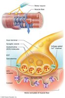
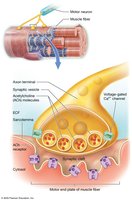
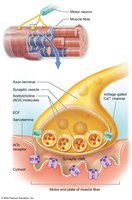
The NMJ is the synapse between a motor neuron and a skeletal muscle fiber. It consists of the axon terminal, synaptic cleft, and motor end plate. The neurotransmitter acetylcholine (ACh) is released from the neuron, binds to receptors on the motor end plate, and initiates an action potential in the muscle fiber.
Skeletal Muscle Contraction: Phases and Mechanisms
Excitation Phase
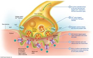
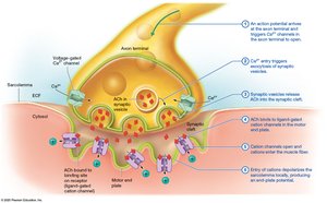
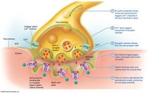
Action potential arrives at the axon terminal, opening voltage-gated calcium channels.
Calcium triggers exocytosis of synaptic vesicles, releasing ACh into the synaptic cleft.
ACh binds to ligand-gated cation channels on the motor end plate, allowing sodium to enter and depolarize the sarcolemma (end-plate potential).
Excitation-Contraction Coupling
End-plate potential triggers an action potential that propagates along the sarcolemma and down T-tubules.
Depolarization of T-tubules opens calcium channels in the SR, releasing calcium into the cytosol.
Contraction Phase: The Crossbridge Cycle
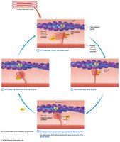
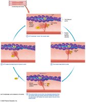
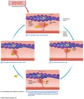
Calcium binds to troponin, causing tropomyosin to move and expose actin's active sites.
Myosin heads bind to actin, forming crossbridges.
ATP hydrolysis "cocks" the myosin head; the power stroke pulls actin toward the M line.
ADP and phosphate are released; ATP binding detaches myosin from actin, and the cycle repeats.
Muscle Relaxation
ACh release stops, and acetylcholinesterase degrades remaining ACh.
Calcium channels in the SR close, and calcium is pumped back into the SR.
Troponin and tropomyosin return to their resting positions, blocking actin's active sites.
Energy Sources for Skeletal Muscle
Immediate: Creatine phosphate donates a phosphate to ADP to form ATP (via creatine kinase).
Glycolytic (Anaerobic): Glycolysis splits glucose into pyruvate, producing ATP without oxygen; if oxygen is low, pyruvate is converted to lactic acid.
Oxidative (Aerobic): Occurs in mitochondria, using oxygen to produce large amounts of ATP from glucose, fatty acids, or amino acids.
Muscle Twitch and Tension Production
A muscle twitch is the response of a muscle fiber to a single action potential. It consists of three phases:
Latent Period: Time for action potential to spread and calcium to be released.
Contraction Period: Crossbridge cycling and tension increase.
Relaxation Period: Calcium is pumped back into the SR, and tension decreases.
Types of Muscle Contractions
Isotonic Concentric: Muscle shortens as it contracts (force > load).
Isotonic Eccentric: Muscle lengthens while contracting (force < load).
Isometric: Muscle length does not change (force = load).
Muscle Fiber Types
Slow-Twitch (Type I): Slow contraction, high endurance, oxidative metabolism, rich in myoglobin (red muscle).
Fast-Twitch (Type II): Fast contraction, fatigue quickly, glycolytic metabolism, less myoglobin (white muscle).
Subtypes: Fast oxidative-glycolytic (FOG) and fast glycolytic (FG).
Motor Units and Recruitment
A motor unit consists of a single motor neuron and all the muscle fibers it innervates. Recruitment of additional motor units increases the force of contraction. Muscle tone is the baseline tension in a muscle, important for posture and readiness.
Physical Training and Muscle Adaptation
Endurance Training: Increases oxidative enzymes, mitochondria, and blood supply; enhances fatigue resistance.
Resistance Training: Increases myofibril number and muscle fiber diameter (hypertrophy); may decrease endurance capacity.
Disuse: Leads to atrophy, decreased oxidative enzymes, and reduced strength/endurance.
Smooth Muscle
Smooth muscle is found in the walls of hollow organs and is responsible for peristalsis, sphincter formation, and regulation of flow. It lacks striations and sarcomeres, and contraction is regulated by calcium binding to calmodulin, which activates myosin light-chain kinase (MLCK).
Cardiac Muscle
Cardiac muscle cells are striated, branched, and connected by intercalated discs. They are autorhythmic, meaning they can generate their own action potentials, and are specialized for continuous, rhythmic contraction to pump blood.
