 Back
BackMuscle Tissue and Physiology: Structured Study Notes
Study Guide - Smart Notes
 Tailored notes based on your materials, expanded with key definitions, examples, and context.
Muscle Tissue and Physiology
Overview of Muscle Tissue
Muscle tissue is essential for movement, posture, joint stabilization, heat generation, and regulation of material flow through hollow organs. It consists of muscle cells (myocytes) and the surrounding endomysium, an extracellular matrix that holds cells together and transmits tension.
Muscle Tension: The force generated by muscle tissue.
Types of Muscle Tissue: Skeletal, Cardiac, and Smooth.
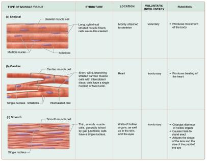
Types of Muscle Tissue
Muscle tissue is classified based on structure, function, and control mechanisms.
Skeletal Muscle: Long, striated, multinucleated cells; voluntary; attached to skeleton; produces movement.
Cardiac Muscle: Short, branched, striated cells with intercalated discs; involuntary; found in heart; produces heartbeat.
Smooth Muscle: Long, flattened cells with single nucleus; involuntary; lines hollow organs; regulates flow and diameter.
Properties of Muscle Cells
Muscle cells possess unique properties that enable their function:
Contractility: Ability to contract and generate force.
Excitability: Ability to respond to stimuli.
Conductivity: Ability to conduct electrical charges.
Distensibility: Ability to be stretched without rupture.
Elasticity: Ability to return to original length after stretching.
Structure of Muscle Cells
Muscle Cell Organelles
Muscle cells share organelles with other cells but have specialized structures:
Sarcoplasm: Muscle cell cytoplasm.
Sarcolemma: Muscle cell plasma membrane.
Myofibrils: Bundles of proteins for contraction; make up 50–80% of cell volume.
Sarcoplasmic Reticulum (SR): Modified smooth ER surrounding myofibrils.
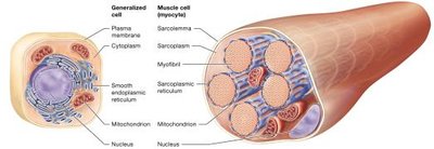
Skeletal Muscle Fiber Structure
Skeletal muscle fibers are long, cylindrical, multinucleated, and striated. They arise from fused embryonic myoblasts.
Myofibrils: Most abundant organelle, surrounded by SR.
Sarcolemma: Forms T-tubules, tunnel-like networks filled with extracellular fluid.
Triad: Combination of a T-tubule and two terminal cisternae.
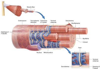
Myofibril and Myofilament Structure
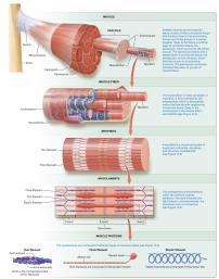
Myofibrils are composed of myofilaments: thick, thin, and elastic filaments.
Thick Filaments: Made of myosin; two heads and a tail.
Thin Filaments: Made of actin (with active sites), tropomyosin (regulatory), troponin (regulatory), and nebulin (structural).
Elastic Filaments: Made of titin; provide elasticity and resist overstretching.
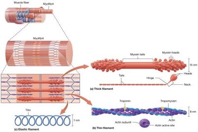
Sarcomere Structure and Bands
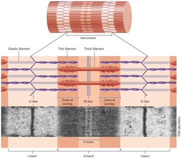
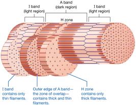
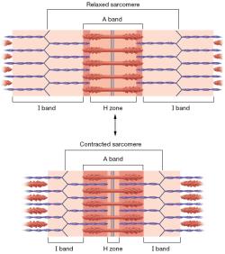
The sarcomere is the functional unit of muscle contraction, defined by distinct bands:
I Band: Light region; only thin filaments.
A Band: Dark region; thick and thin filaments.
H Zone: Middle of A band; only thick filaments.
M Line: Middle of A band; structural proteins.
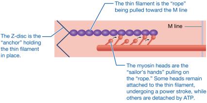
Z-Disc: Anchors thin and elastic filaments; attaches myofibrils.
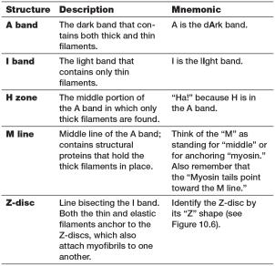
Mnemonic Table for Sarcomere Bands
Structure | Description | Mnemonic |
|---|---|---|
A band | Dark band with thick and thin filaments | A is the dArk band |
I band | Light band with only thin filaments | I is the lIght band |
H zone | Middle of A band; only thick filaments | "Ha!" because H is in the A band |
M line | Middle line; structural proteins | "M" for middle or myosin |
Z-disc | Line bisecting I band; anchors filaments | "Z" shape |
Levels of Organization in Skeletal Muscle
Skeletal muscle is organized hierarchically from muscle to myofibril to myofilament.
Muscle Contraction Mechanisms
Sliding-Filament Mechanism
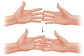
Muscle contraction occurs when thin filaments slide past thick filaments, shortening the sarcomere and generating tension.
I bands and H zone: Narrow during contraction.
A band: Remains unchanged.
Z-discs: Move closer together.
Electrophysiology and Membrane Potential
Membrane Potential and Ion Gradients
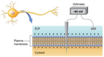
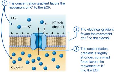
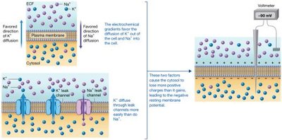
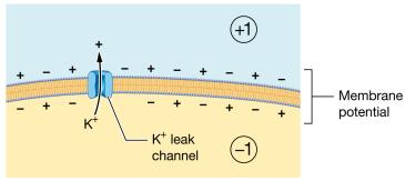
Muscle fibers maintain a resting membrane potential due to ion gradients across the sarcolemma.
Resting Membrane Potential: Typically −90 mV; cell is polarized.
Ion Channels: Leak channels (always open), ligand-gated, and voltage-gated channels.
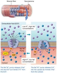
Sodium-Potassium Pump: Moves 3 Na+ out and 2 K+ in, using ATP.
Action Potentials
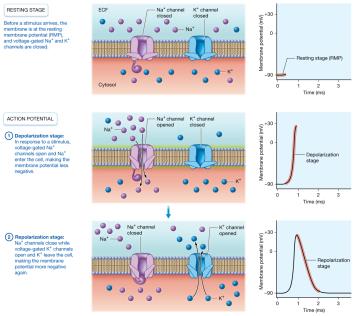
Action potentials are rapid changes in membrane potential, essential for muscle contraction.
Depolarization: Na+ channels open, Na+ enters, membrane becomes less negative.
Repolarization: Na+ channels close, K+ channels open, K+ leaves, membrane returns to negative.
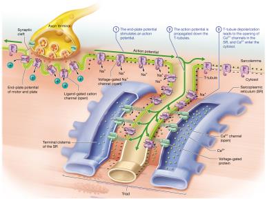
Propagation: Action potential spreads across sarcolemma and T-tubules.
Neuromuscular Junction and Muscle Contraction
Neuromuscular Junction (NMJ)
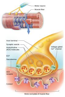
The NMJ is the synapse between a motor neuron and a muscle fiber, facilitating communication via acetylcholine (ACh).
Axon Terminal: Contains synaptic vesicles with ACh.
Synaptic Cleft: Space between neuron and muscle fiber.
Motor End Plate: Region of sarcolemma with ACh receptors.
Phases of Skeletal Muscle Contraction
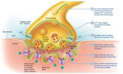
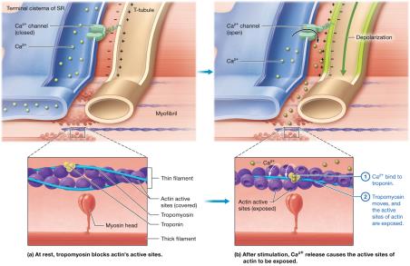
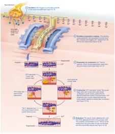
Muscle contraction occurs in three phases: excitation, excitation-contraction coupling, and contraction.
Excitation: ACh released, binds to receptors, depolarizes sarcolemma.
Excitation-Contraction Coupling: Action potential travels down T-tubules, triggers Ca2+ release from SR.
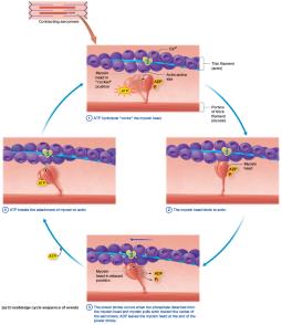
Contraction: Ca2+ binds troponin, tropomyosin moves, myosin binds actin, crossbridge cycle begins.
Muscle Relaxation
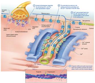
Relaxation occurs when ACh is degraded, Ca2+ is pumped back into SR, and tropomyosin blocks actin sites.
Acetylcholinesterase: Degrades ACh.
Calcium Pumps: Return Ca2+ to SR.
Spasm: Inability to relax; may be caused by dehydration, injury, or overload.
Rigor Mortis
Rigor mortis is the stiffening of muscles after death due to lack of ATP, preventing detachment of myosin from actin.
Energy Sources for Muscle Contraction
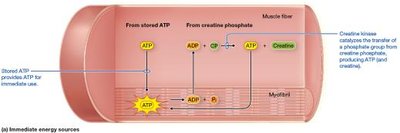
Immediate Energy: Creatine Phosphate
Creatine phosphate regenerates ATP rapidly for short bursts of activity.
Creatine Kinase: Enzyme catalyzing ATP regeneration.
Equation:
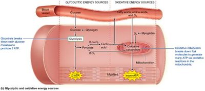
Glycolytic (Anaerobic) Energy
Glycolysis splits glucose into pyruvate, producing ATP without oxygen for moderate-duration activity.
Glycogen: Storage form of glucose in muscle and liver.
Lactic Acid: Produced when oxygen is limited; converted to glucose in liver.
Oxidative (Aerobic) Energy
Oxidative catabolism in mitochondria produces large amounts of ATP using glucose, fatty acids, and amino acids, requiring oxygen.
Myoglobin: Oxygen-binding protein in muscle fibers.
Muscle Tension and Fiber Types
Muscle Twitch and Myogram
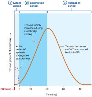
A muscle twitch is the response to a single action potential, recorded as a myogram.
Latent Period: Time for action potential to spread.
Contraction Period: Crossbridge cycling increases tension.
Relaxation Period: Ca2+ pumped back into SR.
Wave Summation and Tetanus
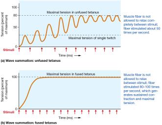
Repeated stimulation increases tension via wave summation, leading to unfused or fused tetanus.
Unfused Tetanus: Partial relaxation between contractions.
Fused Tetanus: No relaxation; sustained contraction.
Length-Tension Relationship
Optimal sarcomere length allows maximal crossbridge formation and tension production.
Overly Shortened: Excessive overlap, less tension.
Overly Stretched: Minimal overlap, less tension.
Optimal Length: Maximal overlap, maximal tension.
Classes of Skeletal Muscle Fibers
Muscle fibers are classified by contraction speed and energy source.
Type I (Slow Oxidative): Slow, fatigue-resistant, red, many mitochondria, extensive blood supply.
Type IIa (Fast Oxidative Glycolytic): Intermediate speed and fatigue, light red, moderate mitochondria.
Type IIx (Fast Glycolytic): Fast, easily fatigued, white, few mitochondria, limited blood supply.
Class | Primary Catabolism | Blood Supply | Mitochondria | Myoglobin | Glycogen | ATPase Activity | Fatigability | Diameter | Color | Example |
|---|---|---|---|---|---|---|---|---|---|---|
Type I | Oxidative | Extensive | Many | Large | Little | Low | Low | Small/intermediate | Red | Standing, sitting |
Type IIa | Oxidative & Glycolytic | Less extensive | Intermediate | Intermediate | Intermediate | High | Intermediate | Large | Light red | Walking, writing |
Type IIx | Glycolytic | Limited | Few | Little | Large | Highest | High | Intermediate | Light pink/white | Heavy lifting, sprinting |
Motor Units and Muscle Contractions
Motor Units
A motor unit consists of a motor neuron and the muscle fibers it innervates. Recruitment of motor units increases contraction force.
Slow Motor Units: Type I fibers.
Fast Motor Units: Type II fibers.
Muscle Tone: Maintains posture and readiness.
Hypotonia: Abnormally low tone.
Hypertonia: Abnormally high tone.
Types of Muscle Contractions
Contractions are classified by changes in muscle length:
Isotonic Concentric: Muscle shortens; force > load.
Isotonic Eccentric: Muscle lengthens; force < load.
Isometric: Muscle length unchanged; force = load.
Adaptations and Fatigue
Physical Training Effects
Endurance Training: Increases oxidative capacity, mitochondria, blood vessels; improves fatigue resistance.
Resistance Training: Increases myofibrils and fiber diameter (hypertrophy); may decrease endurance.
Disuse: Decreases fiber diameter (atrophy), oxidative enzymes, and endurance.
Muscle Fatigue
Fatigue is the inability to maintain exercise intensity, caused by depletion of metabolites, decreased oxygen, accumulation of chemicals, and environmental factors.
Excess Postexercise Oxygen Consumption (EPOC)
Recovery period after exercise involves increased oxygen consumption to restore homeostasis, ion concentrations, and blood pH.
Smooth and Cardiac Muscle
Smooth Muscle
Smooth muscle is found in hollow organs, lacks striations, and contracts via different mechanisms than skeletal muscle.
Functions: Peristalsis, sphincter formation, regulation of flow.
Structure: Actin anchored to dense bodies; lacks troponin.
Contraction: Ca2+ binds calmodulin, activates myosin light-chain kinase, crossbridge cycle ensues.
Types: Single-unit (visceral) and multi-unit.
Cardiac Muscle
Cardiac muscle is striated, contains sarcomeres, T-tubules, and extensive SR. Cells are branched, have one or two nuclei, and are connected by intercalated discs. Pacemaker cells generate action potentials, making cardiac muscle autorhythmic.
Additional info: These notes expand on the original lecture slides and textbook images, providing definitions, examples, and structured tables for clarity. All images included are directly relevant to the adjacent content and reinforce key concepts in muscle tissue and physiology.
