 Back
BackMuscle Tissue: Structure, Organization, and Function
Study Guide - Smart Notes
 Tailored notes based on your materials, expanded with key definitions, examples, and context.
Muscle Tissue
An Introduction to Muscle Tissue
Muscle tissue is a primary tissue in the human body, specialized for contraction and responsible for producing movement. There are three main types of muscle tissue: skeletal muscle, cardiac muscle, and smooth muscle. Each type has unique structural and functional characteristics that enable specific roles in the body.
Functions of Muscles
General Functions
Excitability (Responsiveness): Ability to respond to stimuli.
Contractility: Ability of muscle cells to shorten and generate force.
Extensibility: Ability to be stretched without damage.
Elasticity: Ability to return to original length after stretching.
Functions of Skeletal Muscle
Producing movement by pulling on bones
Maintaining posture and body position
Supporting soft tissues
Guarding body entrances and exits
Maintaining body temperature
Storing nutrients
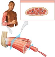
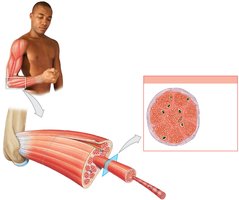
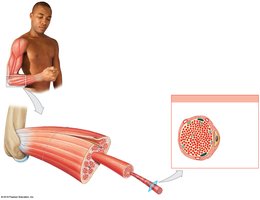
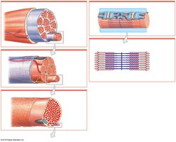
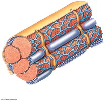
Organization of Skeletal Muscle
Structural Organization
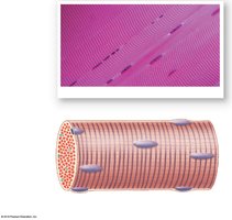
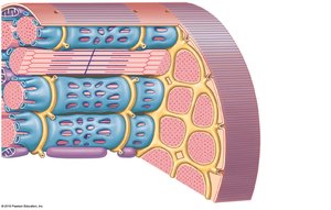
Skeletal muscles are complex organs composed of skeletal muscle tissue, connective tissues, blood vessels, and nerves. The organization of these components allows for efficient force generation and control.
Epimysium: Dense layer of collagen fibers surrounding the entire muscle, separating it from surrounding tissues.
Perimysium: Surrounds bundles of muscle fibers called fascicles; contains blood vessels and nerves.
Endomysium: Surrounds individual muscle fibers; contains capillaries, myosatellite cells (stem cells), and nerve fibers.
The collagen fibers of these connective tissue layers converge to form tendons (bundles) or aponeuroses (sheets) that attach muscles to bones.
Vascular and Neural Supply
Skeletal muscles have extensive vascular networks to deliver oxygen and nutrients and remove wastes.
They contract only when stimulated by the central nervous system (voluntary muscles).
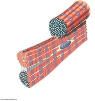
Skeletal Muscle Fibers
Characteristics of Skeletal Muscle Fibers
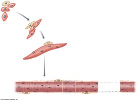
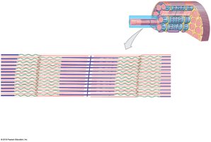
Skeletal muscle fibers are large, multinucleate cells formed by the fusion of embryonic myoblasts. They are also known as striated muscle cells due to their banded appearance.
Sarcolemma: The plasma membrane of a muscle fiber, surrounding the sarcoplasm (cytoplasm).
Transverse tubules (T tubules): Invaginations of the sarcolemma that transmit action potentials into the cell interior, triggering contraction.
Internal Organization
Sarcoplasmic Reticulum (SR): Specialized endoplasmic reticulum that stores and releases calcium ions, essential for muscle contraction.
Triad: Structure formed by a T tubule and two terminal cisternae of the SR.
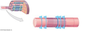
Myofibrils: Cylindrical structures within muscle fibers, composed of bundles of protein filaments (myofilaments).
Myofilaments: Two types—thin filaments (actin) and thick filaments (myosin).
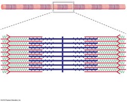
Sarcomeres: The Functional Units of Muscle
Sarcomere Structure
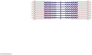
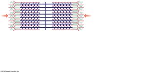
The sarcomere is the smallest functional unit of a muscle fiber. It is defined by the region between two Z lines and is responsible for the striated appearance of skeletal muscle.
A band: Dark region containing thick filaments (myosin) and overlapping thin filaments (actin).
I band: Light region containing only thin filaments.
M line: Center of the A band; stabilizes thick filaments.
H band: Area around the M line with only thick filaments.
Zone of overlap: Region where thick and thin filaments overlap.
Z line: Boundary between adjacent sarcomeres; anchors thin filaments.
Titin: Elastic protein that helps restore sarcomere length after contraction.

Levels of Functional Organization
The organization of skeletal muscle can be summarized as follows:
Level | Surrounded by | Contains |
|---|---|---|
Skeletal Muscle | Epimysium | Muscle fascicles |
Muscle Fascicle | Perimysium | Muscle fibers |
Muscle Fiber | Endomysium | Myofibrils |
Myofibril | Sarcoplasmic reticulum | Sarcomeres |
Sarcomere | — | Thick and thin filaments, titin |
Myofilament Structure
Thin Filaments
F-actin: Twisted strand of two rows of globular G-actin molecules; each G-actin has an active site for myosin binding.
Nebulin: Holds F-actin strands together.
Tropomyosin: Covers active sites on G-actin, preventing actin-myosin interaction.
Troponin: Binds tropomyosin, G-actin, and Ca2+; regulates contraction.
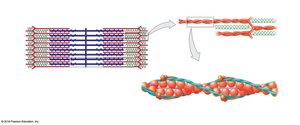
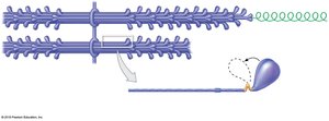
Thick Filaments
Composed of about 300 myosin molecules, each with a tail (binds other myosin) and two heads (bind to actin).
Core of titin helps recoil after stretching.
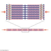
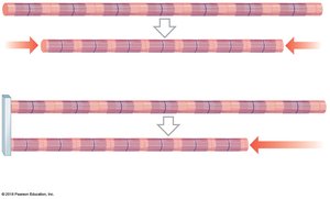
Sliding Filament Theory
Mechanism of Contraction
During muscle contraction, thin filaments slide toward the center of the sarcomere, causing the sarcomere to shorten. The width of the A band remains constant, while the I band and H band narrow.
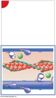
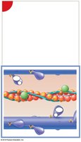
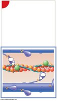
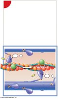
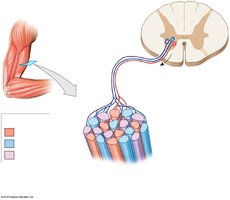
The Neuromuscular Junction (NMJ)
Excitable Membranes and Action Potentials
Muscle fibers and neurons have excitable membranes that can generate action potentials. Skeletal muscle fibers contract in response to stimulation by motor neurons at the NMJ.
Events at the NMJ
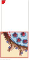
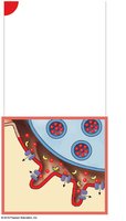
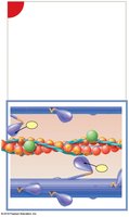
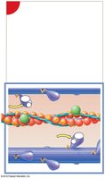
An action potential arrives at the axon terminal of a motor neuron.
Acetylcholine (ACh) is released into the synaptic cleft.
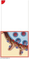
ACh binds to receptors on the motor end plate, opening Na+ channels and depolarizing the sarcolemma.
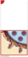
An action potential is generated in the muscle fiber.
ACh is broken down by acetylcholinesterase (AChE), ending the signal.
Excitation-Contraction Coupling
Excitation-contraction coupling links the generation of an action potential in the sarcolemma to the start of a muscle contraction. The action potential travels down T tubules, triggering Ca2+ release from the SR, which binds to troponin and initiates the contraction cycle.
The Contraction Cycle
Active-site exposure (Ca2+ binds troponin, moving tropomyosin)
Cross-bridge formation (myosin binds actin)
Myosin head pivoting (power stroke)
Cross-bridge detachment (ATP binds myosin)
Myosin reactivation (ATP hydrolysis)
Tension Production
Factors Affecting Tension
Number of power strokes performed
Resting length of the fiber at stimulation
Frequency of stimulation
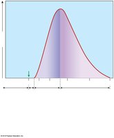
Length-Tension Relationship
Maximum tension is produced when the maximum number of cross-bridges can form, which occurs at an optimal sarcomere length.
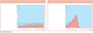
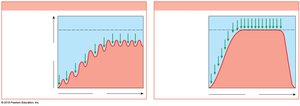
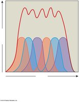
Frequency of Stimulation
Twitch: Single contraction from one stimulus (three phases: latent, contraction, relaxation)
Treppe: Gradual increase in tension with repeated stimulation after relaxation
Wave Summation: Increased tension from repeated stimuli before relaxation ends
Tetanus: Maximum tension (incomplete or complete)
Muscle Contractions
Motor Units and Recruitment
Motor unit: A motor neuron and all the muscle fibers it controls.
Recruitment: Increasing the number of active motor units for greater tension.
Muscle tone: Normal tension and firmness at rest, important for posture and stability.
Types of Muscle Contractions
Isotonic: Muscle changes length (concentric: shortens; eccentric: lengthens)
Isometric: Muscle develops tension but does not change length
Energy for Muscle Contraction
ATP and Muscle Metabolism
ATP is the direct energy source for contraction.
ATP is generated by:
Direct phosphorylation (creatine phosphate)
Anaerobic metabolism (glycolysis)
Aerobic metabolism (citric acid cycle and electron transport chain)
Muscles store glycogen and creatine phosphate for rapid ATP production.
Oxygen Debt and Recovery
After exercise, extra oxygen is required to restore normal conditions (EPOC).
Lactate is recycled in the liver (Cori cycle).
Muscle Performance
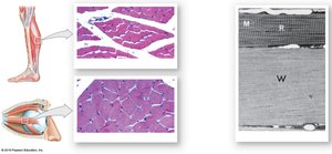
Types of Skeletal Muscle Fibers
Fast fibers: Large, powerful, fatigue quickly, few mitochondria, pale color.
Slow fibers: Small, fatigue-resistant, many mitochondria, high myoglobin, dark color.
Intermediate fibers: Mid-sized, moderate endurance, little myoglobin.
Muscle Hypertrophy and Atrophy
Hypertrophy: Increase in muscle size due to training.
Atrophy: Decrease in muscle size due to inactivity.
Aging and Muscle Fatigue
Muscle fibers decrease in size and elasticity with age.
Fatigue results from depletion of energy reserves, pH changes, and structural damage.
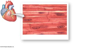
Cardiac Muscle Tissue
Structure and Function
Found only in the heart; striated, small, branched cells with a single nucleus.
Connected by intercalated discs (gap junctions and desmosomes) for synchronized contraction.
Automaticity: Can contract without neural stimulation (pacemaker cells).
Smooth Muscle Tissue
Structure and Function
Found in walls of hollow organs, blood vessels, and other systems.
Spindle-shaped cells, single central nucleus, nonstriated.
No T tubules or sarcomeres; thin filaments attached to dense bodies.
Can contract over a wide range of lengths (plasticity).
Control of Contraction
Excitation-contraction coupling involves Ca2+ binding to calmodulin.
Multiunit smooth muscle: Each cell innervated individually.
Visceral smooth muscle: Cells connected, contract as a unit, controlled by pacesetter cells.
Smooth Muscle Tone
Maintains a normal background level of activity, modulated by neural, hormonal, or chemical factors.
