 Back
BackMuscles of the Lower Limb: Structure, Function, Attachments, and Innervation
Study Guide - Smart Notes
 Tailored notes based on your materials, expanded with key definitions, examples, and context.
Muscles of the Lower Limb
Introduction
The muscles of the lower limb are essential for stabilizing the pelvic girdle and facilitating movement such as standing, walking, and running. These muscles are organized into compartments and act on the hip, thigh, leg, and foot, each with specific actions, attachments, and innervation.
Muscles That Act on the Hip and Femur
Gluteal and Lateral Hip Muscles
The gluteal muscles are among the largest and most powerful in the body, responsible for movements at the hip joint. The gluteus maximus extends the thigh, while the gluteus medius and gluteus minimus abduct the thigh. The tensor fascia latae assists in flexion and abduction and stabilizes the knee via the iliotibial tract. Deep lateral rotators, such as the piriformis and obturator muscles, rotate the femur laterally.
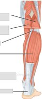
Iliopsoas: Flexes and laterally rotates the thigh; flexes the torso.
Gluteus maximus: Extends the thigh.
Gluteus medius/minimus: Abduct the thigh.
Tensor fascia latae: Flexes and abducts the thigh; stabilizes the knee.
Adductors (longus, brevis, magnus): Adduct and rotate the thigh; flex or extend depending on the muscle.
Pectineus: Adducts and flexes the femur at the hip.
Table: Muscles of the Hip and Thigh
Muscle Name | Primary Action | Origin | Insertion | Innervation |
|---|---|---|---|---|
Iliopsoas | Flex, laterally rotate thigh; flex torso | Lumbar vertebrae, iliac fossa | Lesser trochanter | Femoral nerve |
Gluteus maximus | Extend thigh | Dorsal ilium, sacrum, coccyx | Gluteal tuberosity, IT tract | Inferior gluteal nerve |
Gluteus medius | Abduct thigh | Superior lateral ilium | Greater trochanter | Superior gluteal nerve |
Adductor longus | Adduct, medially rotate, flex thigh | Pubis | Linea aspera | Obturator nerve |
Pectineus | Adduct, flex, medially rotate thigh | Pectineal line of pubis | Lesser trochanter, linea aspera | Femoral nerve |
Thigh Muscles That Act on the Femur, Tibia, and Fibula
Anterior, Medial, and Posterior Compartments
The thigh is divided into compartments by deep fascia. The anterior compartment contains the quadriceps femoris group, which extends the knee and stabilizes the joint. The medial compartment adducts the femur, while the posterior compartment (hamstrings) flexes the knee and extends the thigh.
Quadriceps femoris: Four muscles (rectus femoris, vastus lateralis, vastus medialis, vastus intermedius) that extend the knee.
Sartorius: Flexes, abducts, and laterally rotates the thigh; flexes the knee.
Hamstrings (biceps femoris, semitendinosus, semimembranosus): Flex the knee and extend the thigh.
Gracilis: Adducts the thigh and flexes the knee.
Table: Thigh Muscles that Move the Femur, Tibia, and Fibula
Muscle Name | Primary Action | Origin | Insertion | Innervation |
|---|---|---|---|---|
Gracilis | Adduct thigh, flex knee | Pubis, ischial ramus | Medial tibia | Obturator nerve |
Sartorius | Flex, abduct, laterally rotate thigh; flex knee | ASIS | Medial proximal tibia | Femoral nerve |
Rectus femoris | Extend knee, flex thigh | AIIS, acetabulum | Patella, tibial tuberosity | Femoral nerve |
Biceps femoris | Extend, laterally rotate thigh; flex knee | Ischial tuberosity, femur | Fibular head, lateral tibia | Sciatic nerve |
Muscles of the Lower Leg
Anterior, Lateral, and Posterior Compartments
The lower leg muscles are divided into three compartments. The anterior compartment dorsiflexes the foot and extends the toes. The lateral compartment everts and plantar flexes the foot. The posterior compartment (superficial and deep) plantar flexes the foot and flexes the toes.
Tibialis anterior: Dorsiflexes and inverts the foot.
Extensor digitorum longus: Extends toes, dorsiflexes foot.
Fibularis longus/brevis: Plantar flex and evert foot.
Gastrocnemius: Plantar flexes foot, flexes knee.
Soleus: Plantar flexes foot.
Flexor digitorum longus: Plantar flexes and inverts foot; flexes toes.
Flexor hallucis longus: Plantar flexes foot; flexes big toe.
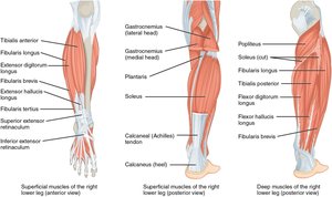
Table: Muscles that Move the Feet and Toes
Muscle Name | Primary Action | Origin | Insertion | Innervation |
|---|---|---|---|---|
Gastrocnemius | Plantar flex foot; flex knee | Femoral condyles | Posterior calcaneus | Tibial nerve |
Soleus | Plantar flex foot | Tibia, fibula | Posterior calcaneus | Tibial nerve |
Tibialis anterior | Dorsiflex, invert foot | Lateral tibia | Medial cuneiform, 1st metatarsal | Deep fibular nerve |
Fibularis longus | Plantar flex, evert foot | Lateral fibula | 1st metatarsal, medial cuneiform | Superficial fibular nerve |
Intrinsic Muscles of the Foot
Structure and Function
The intrinsic muscles of the foot originate and insert within the foot, supporting the arch and enabling toe movements. The plantar aponeurosis provides principal support for the longitudinal arch. The dorsal group includes the extensor digitorum brevis, while the plantar group consists of four layers of muscles.
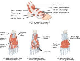
Muscle Actions, Attachments, and Innervation
Muscle Actions
Each muscle has specific actions, such as flexion, extension, abduction, adduction, and rotation of the lower limb at various joints.
For example, the gastrocnemius plantar flexes the foot and flexes the knee, while the tibialis anterior dorsiflexes and inverts the foot.
Muscle Attachments
Muscle origins and insertions are critical for understanding their actions. For example, the rectus femoris originates from the anterior inferior iliac spine and inserts on the patella and tibial tuberosity.
The fibularis longus originates from the upper lateral fibula and inserts on the first metatarsal and medial cuneiform.
Muscle Innervation
Femoral nerve: Innervates iliopsoas, sartorius, quadriceps femoris, pectineus.
Obturator nerve: Innervates adductor longus, adductor brevis, adductor magnus, gracilis.
Tibial nerve: Innervates gastrocnemius, soleus, plantaris, tibialis posterior, flexor digitorum longus, flexor hallucis longus.
Sciatic nerve: Innervates biceps femoris, semitendinosus, semimembranosus.
Summary Table: Major Lower Limb Muscles, Actions, Attachments, and Innervation
Muscle | Action | Origin | Insertion | Nerve |
|---|---|---|---|---|
Gluteus maximus | Extend thigh | Ilium, sacrum, coccyx | Gluteal tuberosity, IT tract | Inferior gluteal |
Rectus femoris | Extend knee, flex thigh | AIIS | Patella, tibial tuberosity | Femoral |
Biceps femoris | Flex knee, extend thigh | Ischial tuberosity, femur | Fibular head, lateral tibia | Sciatic |
Gastrocnemius | Plantar flex foot, flex knee | Femoral condyles | Calcaneus | Tibial |
Tibialis anterior | Dorsiflex, invert foot | Lateral tibia | Medial cuneiform, 1st metatarsal | Deep fibular |
Key Concepts and Applications
Understanding muscle compartments and their innervation is essential for diagnosing nerve injuries and movement disorders.
Knowledge of muscle attachments helps explain the mechanics of movement and common injuries, such as strains and tears.
Precise terminology is required for describing muscle actions, especially in clinical and academic settings.
Additional info: The tables and images provided are derived from OpenStax Anatomy & Physiology, ensuring alignment with standard college-level curriculum.
