 Back
BackMuscular Tissue: Structure, Function, and Microscopic Anatomy
Study Guide - Smart Notes
 Tailored notes based on your materials, expanded with key definitions, examples, and context.
Muscular Tissue
Types of Muscle Tissue
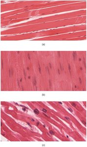
Muscle tissue is classified into three main types, each with distinct structural and functional characteristics. These types are essential for various physiological processes in the human body.
Skeletal muscle: Voluntary, striated muscle attached to bones, responsible for body movements.
Cardiac muscle: Involuntary, striated muscle found only in the heart, responsible for pumping blood.
Smooth muscle: Involuntary, non-striated muscle found in walls of hollow organs (e.g., intestines, blood vessels).
Muscle cells are also known as myocytes, muscle fibers, or myofibers.
Functions of Muscular Tissue
Muscular tissue performs several vital functions in the body:
Producing body movements: Facilitates locomotion and manipulation of the environment.
Stabilizing body positions: Maintains posture and joint stability.
Storing & mobilizing substances: Controls passage of substances via sphincters and peristalsis; heart pumps blood.
Generating heat: Muscle metabolism produces heat (thermogenesis), such as shivering.
Properties of Muscle Tissue
Muscle tissue exhibits four key properties that enable its function:
Electrical excitability: Ability to respond to stimuli by generating action potentials.
Contractility: Ability to shorten forcefully when stimulated.
Extensibility: Ability to stretch without damage.
Elasticity: Ability to return to original shape after contraction or extension.
Structure of Skeletal Muscle Tissue
Connective Tissue Components
Skeletal muscle is organized and supported by several connective tissue layers:
Hypodermis: Adipose and areolar connective tissue connecting skin to fascia.
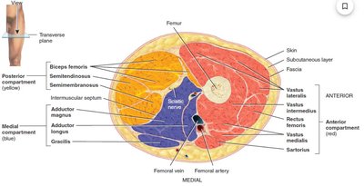
Fascia: Dense irregular connective tissue that groups muscles with similar functions and separates them into compartments.
Mysia: Extensions of fascia, including:
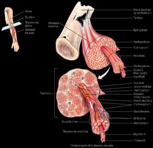
Epimysium: Surrounds entire muscle.
Perimysium: Surrounds muscle fascicles (bundles of fibers).
Endomysium: Surrounds individual muscle fibers.
These layers allow free movement, carry nerves and blood vessels, and fill spaces between muscles.
Tendons, Nerve, and Blood Supply
Tendons are formed by the continuation of epimysium and perimysium beyond the muscle fibers, connecting muscle to bone. Each muscle is supplied by nerves, arteries, and veins, ensuring proper function and nourishment.
Muscle Fiber Organization
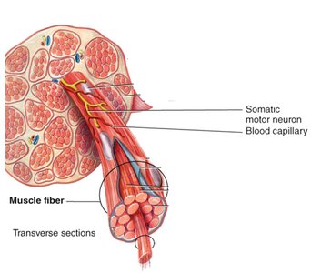
Muscle fibers are the basic cellular units of skeletal muscle. They are organized into fascicles, which are further grouped to form the entire muscle.
Muscle fiber: Individual cell, separated by endomysium.
Muscle fascicle: Bundle of muscle fibers, separated by perimysium.
Muscle: Bundle of fascicles, separated by epimysium.
Analogy: Like pencils in a box, each pencil represents a myofibril, and the box is the muscle fiber.

Microscopic Anatomy of a Skeletal Muscle Fiber
Cellular Components
Skeletal muscle fibers have specialized structures for contraction and energy storage:
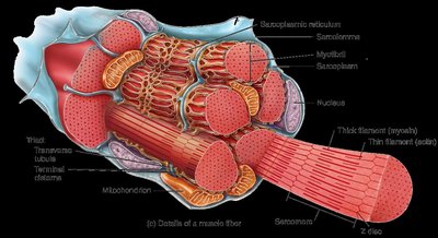
Sarcolemma: Plasma membrane of the muscle fiber, with transverse tubules (T-tubules) for signal transmission.
Sarcoplasm: Cytoplasm containing myoglobin for oxygen storage.
Myofibrils: Cylindrical structures composed of myofilaments, responsible for contraction.
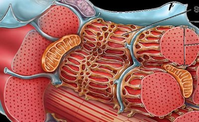
Sarcoplasmic reticulum: Specialized endoplasmic reticulum storing calcium ions, with terminal cisterns.
Muscle Fiber Development and Repair
Skeletal muscle fibers are formed by the fusion of embryonic myoblasts. Although mature fibers are post-mitotic, satellite cells assist in repair. Smooth muscle repairs more readily than skeletal or cardiac muscle. Muscle can undergo hypertrophy (increase in size), atrophy (decrease in size), and fibrosis (replacement with scar tissue).
Filaments and the Sarcomere
Myofilaments and Sarcomere Structure
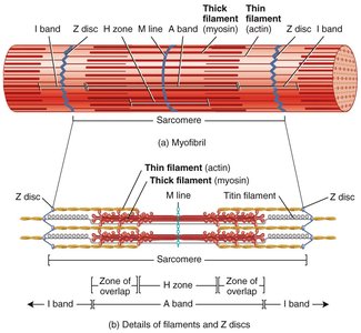
Myofibrils are composed of protein myofilaments:
Thick filaments: Made of myosin.
Thin filaments: Made mostly of actin.
Myofilaments are organized into sarcomeres, the basic functional units of muscle contraction.
Sarcomere Bands and Lines
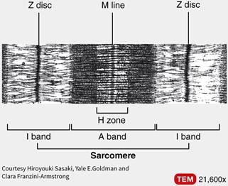
Sarcomeres display specific patterns:
Z disc/line: Boundary of the sarcomere.
A band: Region containing thick filaments.
I band: Region containing only thin filaments.
H zone: Central region with only thick filaments.
M line: Center of the sarcomere, supporting thick filaments.
Muscle Proteins
Types of Muscle Proteins
Muscle contraction and structure depend on three categories of proteins:
Contractile proteins: Myosin and Actin (generate force).
Regulatory proteins: Troponin and Tropomyosin (control contraction).
Structural proteins: Titin, Nebulin, Alpha-actin, Myomesin, Dystrophin (maintain alignment and elasticity).
Muscular Dystrophy
Overview and Pathology
Muscular dystrophy is a group of hereditary diseases characterized by degeneration and weakening of skeletal muscles, replaced by fat and fibrous tissue. Duchenne muscular dystrophy is the most common form, caused by a mutation in the gene for dystrophin, affecting boys and leading to early mortality.
Dystrophin mutation: Disrupts linkage between actin and sarcolemma, causing membrane damage, necrosis, and scar tissue formation.
Example: Duchenne muscular dystrophy is diagnosed between ages 2-10 and is incurable.
Summary Table: Muscle Tissue Types
Type | Location | Control | Appearance | Function |
|---|---|---|---|---|
Skeletal | Attached to bones | Voluntary | Striated | Movement, posture |
Cardiac | Heart | Involuntary | Striated | Pumping blood |
Smooth | Walls of hollow organs | Involuntary | Non-striated | Movement of substances |
Key Equations
Muscle contraction involves the generation of force, which can be described by the sliding filament theory:
ATP hydrolysis provides energy for contraction:
Additional info: Academic context was added to clarify muscle fiber organization, sarcomere structure, and muscle protein functions.
