Back
BackNervous System III: Senses – Structured Study Notes
Study Guide - Smart Notes
 Tailored notes based on your materials, expanded with key definitions, examples, and context.
Nervous System III: Senses
General Characteristics of Sensory Function
The senses are essential for maintaining homeostasis by providing information about both the external environment and internal conditions. Sensory function is divided into general senses and special senses, each with distinct anatomical distributions and functions.
General senses: Receptors are widely distributed throughout the body (skin, organs, joints).
Special senses: Specialized receptors confined to structures in the head (eyes, ears, nose, mouth).
Sensory receptors: Collect information from the environment and relay it to the CNS via sensory neurons.
Pathways for Sensory Information
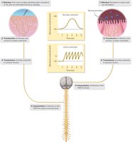
Sensory information follows a specific pathway from the receptor to the CNS:
Stimulation: A stimulus activates sensory receptors.
Transduction: The stimulus is converted into graded receptor potentials.
Transmission: Receptor potentials may trigger action potentials, which are conducted along sensory neurons to the CNS.
Interpretation: The CNS (brain or spinal cord) interprets the sensory information.
Receptors, Sensation, and Perception
Sensory receptors are specialized to respond to specific stimuli, allowing the body to interpret sensory events. Sensation and perception are distinct processes:
Sensation: The brain becomes aware of a sensory event (e.g., pain).
Perception: The brain interprets the sensory impulses (e.g., realizing pain is from stepping on a tack).
Projection: The cerebral cortex projects the sensation back to the apparent source, allowing localization of the stimulus.
Types of Sensory Receptors
There are five main types of sensory receptors:
Chemoreceptors: Respond to chemical changes (smell, taste, oxygen concentration).
Pain receptors (nociceptors): Respond to tissue damage (mechanical, electrical, thermal energy).
Thermoreceptors: Respond to moderate temperature changes.
Mechanoreceptors: Respond to mechanical forces (touch, tension, blood pressure, stretch).
Photoreceptors: Respond to light (eyes).
Sensory Impulses
Sensory receptors may be neuron endings or cells near neuron extensions. Stimulation causes a local change in membrane potential, which may generate an action potential if the receptor is part of a neuron. Peripheral nerves transmit impulses to the CNS for analysis and interpretation.
Sensory Adaptation
Sensory adaptation is the ability to ignore unimportant or continuous stimuli. It involves decreased response from receptors or CNS pathways, requiring a stronger stimulus to trigger impulses. Thermoreceptors and olfactory receptors are especially adept at adaptation.
General Senses
Classification of General Senses
General senses are associated with small, widespread sensory receptors in the skin, muscles, joints, and viscera. They are divided into three groups:
Exteroceptive senses: Associated with the body surface (touch, pressure, temperature, pain).
Interoceptive (visceroceptive) senses: Associated with changes in the viscera (e.g., blood pressure).
Proprioceptive senses: Associated with changes in muscles, tendons, and joints (body position).
Touch and Pressure Senses
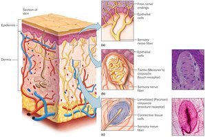
Three types of mechanoreceptors respond to touch and pressure:
Free nerve endings: Common in epithelial tissues; sense itching and other sensations.
Tactile (Meissner’s) corpuscles: Abundant in hairless skin and lips; detect fine touch and texture.
Lamellated (Pacinian) corpuscles: Found in deeper tissues, tendons, ligaments; detect heavy pressure and vibrations.
Temperature Senses
Temperature receptors (thermoreceptors) are free nerve endings in the skin. There are two types:
Warm receptors: Sensitive to temperatures above 25°C, unresponsive above 45°C.
Cold receptors: Sensitive to temperatures between 10°C and 20°C.
Pain receptors: Respond to extreme temperatures below 10°C or above 45°C.
Sense of Pain
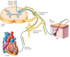
Pain receptors (nociceptors) are free nerve endings widely distributed except in the brain. They respond to tissue damage, chemicals, mechanical forces, temperature extremes, and oxygen deficiency. Pain receptors adapt very little.
Visceral Pain and Referred Pain
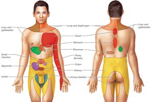
Pain receptors in viscera are the only ones whose stimulation produces sensations. Visceral pain may be felt as coming from another part of the body, a phenomenon known as referred pain. This occurs due to common nerve pathways in the CNS.
Pain Pathways
There are two types of fibers conducting pain impulses:
Fast pain (A-delta) fibers: Myelinated, conduct rapidly, associated with sharp, localized pain.
Slow pain (C) fibers: Unmyelinated, conduct slowly, associated with dull, aching pain.
Regulation of Pain Pathways
The thalamus begins the sensation of pain, while the cerebral cortex judges intensity and location. Emotional responses involve the limbic system. Pain-inhibiting substances include enkephalins, serotonin, and endorphins.
Proprioception
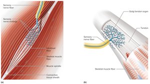
Proprioceptors are mechanoreceptors that provide information about body position and muscle tension. Main types include:
Lamellated (Pacinian) corpuscles: Pressure receptors in joints.
Muscle spindles: Stretch receptors in skeletal muscles; initiate stretch reflexes.
Golgi tendon organs: Stretch receptors in tendons; stimulate reflexes that oppose stretch reflexes.
Visceral Senses
Visceral senses have receptors in internal organs, such as lamellated corpuscles and free nerve endings. They convey information about fullness, discomfort, and pain from internal organs.
Special Senses
Overview of Special Senses
Special senses have sensory receptors within large, complex organs in the head:
Smell: Olfactory organs in nasal cavity.
Taste: Taste buds in oral cavity.
Hearing and equilibrium: Inner ears.
Sight: Eyes.
Sense of Smell: Olfaction
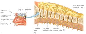
Olfactory receptors are chemoreceptors that respond to chemicals dissolved in liquids. Olfactory organs contain receptor cells and supporting epithelial cells, located in the upper parts of the nasal cavity. Odorants bind to membrane receptors, resulting in depolarization and action potentials.
Olfactory Pathways
Olfactory impulses travel through the cribriform plate to olfactory bulbs, then to olfactory tracts, limbic system, and olfactory cortex. The limbic system provides emotional responses to odors.
Olfactory Stimulation
Each olfactory receptor cell contains one type of membrane protein, which can bind several odorants. Olfactory adaptation is rapid, and receptor neurons are regularly replaced.
Sense of Taste: Gustation
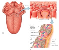
Taste buds are organs of taste, located on papillae of the tongue, roof of mouth, cheeks, and pharynx. Taste cells are modified epithelial cells with microvilli (taste hairs) that protrude through taste pores.
Taste Sensations
There are five primary taste sensations:
Sweet: Stimulated by carbohydrates.
Sour: Stimulated by acids.
Salty: Stimulated by salts.
Bitter: Stimulated by organic compounds, Mg and Ca salts.
Umami: Stimulated by amino acids, MSG.
Taste Pathways
Taste impulses travel via the facial, glossopharyngeal, and vagus nerves to the medulla oblongata, thalamus, and gustatory cortex in the insula.
Sense of Hearing
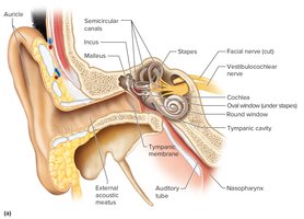
The ear is the organ of hearing and equilibrium, divided into three sections:
Outer ear: Auricle, external acoustic meatus, tympanic membrane.
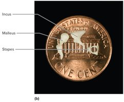
Middle ear: Tympanic cavity, auditory ossicles (malleus, incus, stapes), oval window.
Inner ear: Osseous and membranous labyrinths, cochlea, semicircular canals, vestibule.
Middle Ear: Tympanic Reflex
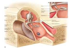
Tympanic reflex involves muscle contractions during loud sounds to protect hearing receptors. Muscles include tensor tympani and stapedius.
Inner (Internal) Ear
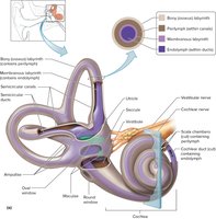
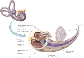
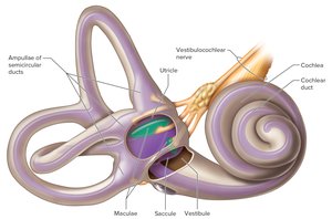
The inner ear contains the cochlea (hearing), semicircular canals (dynamic equilibrium), and vestibule (static equilibrium). It consists of bony and membranous labyrinths filled with perilymph and endolymph.
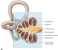
Cochlea and Hearing
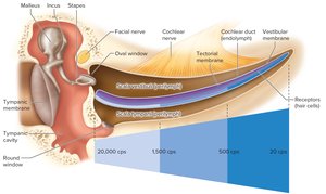
The cochlea is a spiral tube with three compartments: scala vestibuli, scala tympani, and cochlear duct. The cochlear duct contains the spiral organ (organ of Corti), the receptor organ for hearing.
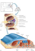
Spiral Organ (Organ of Corti)
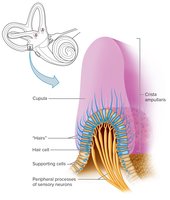
The spiral organ sits on the basilar membrane and contains hair cells with stereocilia. Sound vibrations cause stereocilia to bend against the tectorial membrane, generating nerve impulses.
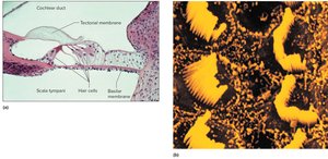
Auditory Pathways
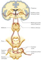
Auditory impulses travel from the cochlear branch of the vestibulocochlear nerve to the medulla oblongata, midbrain, thalamus, and auditory cortex in the temporal lobe.
Sense of Equilibrium
Equilibrium is derived from static and dynamic senses:
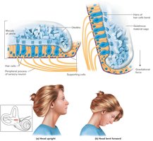
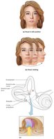
Static equilibrium: Senses head position when not moving; receptors in vestibule (utricle and saccule).
Dynamic equilibrium: Senses rotation and movement; receptors in semicircular canals (crista ampullaris).
Sense of Sight: Vision
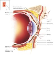
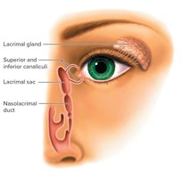
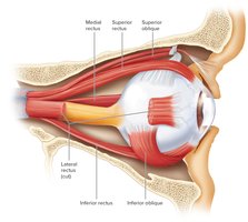
Visual receptors are found in the eye, with accessory organs including eyelids, eyelashes, lacrimal apparatus, and extrinsic eye muscles.
Structure of the Eye
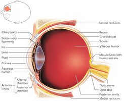
The eye is a hollow, spherical organ with three layers:
Outer (fibrous) tunic: Cornea and sclera.
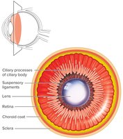
Middle (vascular) tunic: Choroid coat, ciliary body, iris.
Inner (nervous) tunic: Retina.
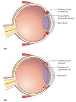
The Iris and Aqueous Humor
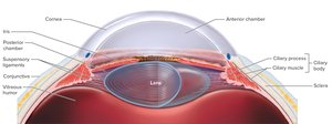
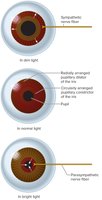
The iris controls light entry, and the aqueous humor provides nutrients and maintains shape. The pupil dilates or constricts in response to light.
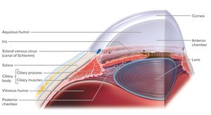
Posterior Cavity and Retina
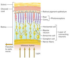
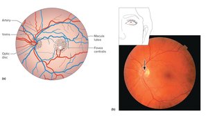
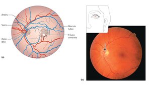
The posterior cavity contains vitreous humor, which maintains eye shape. The retina contains photoreceptors (rods and cones), macula lutea, fovea centralis, and optic disc.
Light Refraction and Lenses
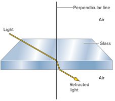
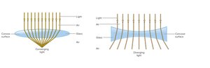
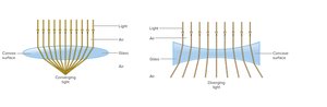
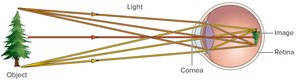
Refraction is the bending of light as it passes between media of different densities. The cornea and lens focus light on the retina, forming an inverted image corrected by the visual cortex.
Refraction Disorders
Common refraction disorders include:
Presbyopia: Age-related farsightedness due to loss of lens elasticity.
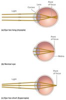
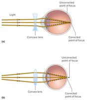
Myopia: Nearsightedness; eyeball too long, corrected by concave lens.
Hyperopia: Farsightedness; eyeball too short, corrected by convex lens.
Astigmatism: Defect in curvature of cornea or lens; corrected with glasses.
Photoreceptors
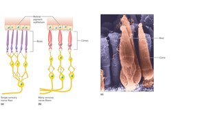
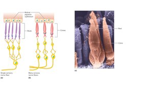
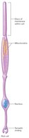
Photoreceptors are modified neurons in the retina:
Rods: Sensitive to dim light, provide vision in shades of gray.
Cones: Sensitive to bright light, provide sharp and color vision.
Visual Pigments
Rods contain rhodopsin, which decomposes in light to trigger nerve impulses. Cones contain iodopsins (erythrolabe, chlorolabe, cyanolabe), each sensitive to different wavelengths for color vision.
Stereoscopic Vision
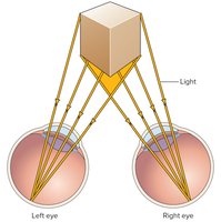
Stereoscopic vision allows perception of depth, height, and width due to the distance between pupils and formation of two slightly different retinal images.
Visual Pathways
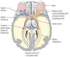
Visual impulses proceed from ganglion cells of the retina to the optic nerve, optic chiasma, optic tracts, thalamus, optic radiations, and visual cortex in the occipital lobe.