 Back
BackNeurons: Cellular and Network Properties – Study Notes
Study Guide - Smart Notes
 Tailored notes based on your materials, expanded with key definitions, examples, and context.
Neurons: Cellular and Network Properties
Electrical Signals in Neurons
Neurons communicate through electrical signals generated by the movement of ions across their membranes. The membrane potential is determined by the distribution and movement of ions such as potassium (K+), sodium (Na+), and chloride (Cl–).
Nernst Equation: Predicts the equilibrium potential for a single ion type if the membrane is permeable only to that ion.
Goldman-Hodgkin-Katz (GHK) Equation: Calculates the membrane potential considering the permeability and concentration gradients of multiple ions.
Resting Membrane Potential: Primarily determined by the K+ gradient and the cell's permeability to K+, Na+, and Cl–.
Key Equations:
Nernst Equation:
GHK Equation:
Ion Concentrations and Equilibrium Potentials
The equilibrium potential for each ion is determined by its concentration gradient across the membrane. Typical values for major ions are as follows:
Ion | Extracellular Fluid (mM) | Intracellular Fluid (mM) | Eion at 37°C (mV) |
|---|---|---|---|
K+ | 5 | 150 | –90 |
Na+ | 145 | 15 | +60 |
Cl– | 108 | 10 | –63 |
Ca2+ | 1 | 0.0001 | See Concept Check |
Example: K+ tends to move out of the cell, while Na+ and Cl– tend to move in.
Ion Movement and Electrical Signals
Changes in membrane permeability to ions generate electrical signals. Opening or closing ion channels alters the flow of ions, leading to depolarization (membrane potential becomes less negative) or hyperpolarization (more negative).
Depolarization: Membrane potential becomes less negative (e.g., Na+ entry).
Hyperpolarization: Membrane potential becomes more negative (e.g., K+ exit or Cl– entry).

Gated Ion Channels
Ion channels are classified by their gating mechanisms:
Mechanically Gated: Open in response to physical deformation.
Chemically Gated: Open in response to ligand binding.
Voltage-Gated: Open in response to changes in membrane potential.
Each channel type has a specific threshold voltage and activation/inactivation kinetics.
Ohm’s Law in Neurons
Current flow in neurons follows Ohm’s Law:
Where V is voltage, I is current, and R is resistance.
Low resistance increases current flow; high resistance decreases it.
Types of Electrical Signals: Graded and Action Potentials
Neurons use two main types of electrical signals:
Graded Potentials: Variable strength, short-distance signals, can be depolarizing or hyperpolarizing.
Action Potentials: All-or-none, large depolarizations, rapid long-distance signaling.
Type of Signal | Location | Channels Involved | Ions | Strength | Initiation | Unique Features |
|---|---|---|---|---|---|---|
Graded | Dendrites, cell body | Mechanically, chemically, voltage-gated | Na+, K+, Ca2+ | Variable, can sum | Entry of ions | No threshold, can sum |
Action | Axon hillock, axon | Voltage-gated | Na+, K+ | All-or-none | Above-threshold graded potential | Threshold required, refractory period |
Graded Potentials
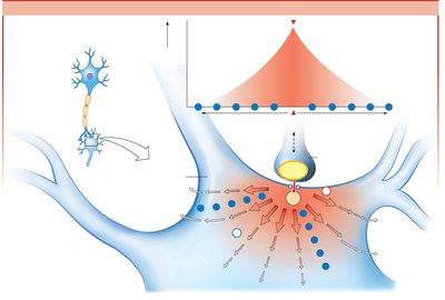
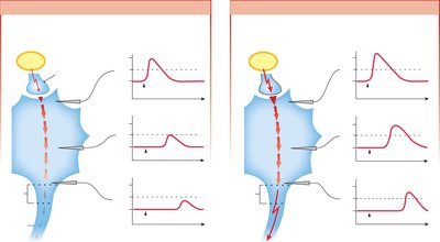
Graded potentials decrease in strength as they spread from the point of origin due to current leak and cytoplasmic resistance. If strong enough, they reach the trigger zone and may initiate an action potential.
Excitatory: Depolarize the membrane, increasing likelihood of action potential.
Inhibitory: Hyperpolarize the membrane, decreasing likelihood of action potential.
Action Potentials
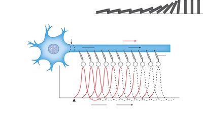
Action potentials are rapid, large depolarizations that travel along the axon without losing strength. They are initiated when a graded potential reaches threshold at the trigger zone.
Rising Phase: Voltage-gated Na+ channels open, Na+ enters, depolarizing the cell.
Falling Phase: Na+ channels inactivate, K+ channels open, K+ exits, repolarizing and hyperpolarizing the cell.
Return to Resting Potential: K+ channels close, membrane returns to resting state.

Voltage-Gated Na+ Channels and Refractory Periods
These channels have two gates (activation and inactivation) that regulate Na+ flow. The absolute refractory period (no new action potential possible) is followed by a relative refractory period (requires stronger stimulus).
Absolute Refractory Period: Ensures one-way propagation and limits firing rate.
Relative Refractory Period: Some Na+ channels reset, but K+ channels remain open.


Conduction of Action Potentials
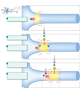
Action potentials propagate by local current flow, with positive charge spreading to adjacent regions, triggering new action potentials. The refractory period prevents backward conduction.
Speed of Action Potential Conduction
Conduction velocity depends on axon diameter and myelination:
Larger Diameter: Less resistance, faster conduction.
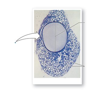
Myelination: Insulates axon, allowing saltatory conduction between nodes of Ranvier.
Demyelinating Diseases: Such as multiple sclerosis, slow or block conduction.

Chemical Factors Affecting Electrical Activity
Certain chemicals and changes in extracellular ion concentrations (especially K+ and Ca2+) can alter neuronal excitability:
Hyperkalemia: Increases excitability by bringing membrane closer to threshold.
Hypokalemia: Decreases excitability by hyperpolarizing the membrane.

Synaptic Transmission and Neurotransmitter Release
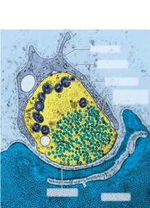
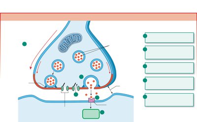
Neurons communicate at synapses via neurotransmitter release:
Action potential arrives at axon terminal.
Voltage-gated Ca2+ channels open, Ca2+ enters.
Ca2+ triggers exocytosis of synaptic vesicles, releasing neurotransmitter.
Neurotransmitter diffuses across synaptic cleft, binds to receptors on postsynaptic cell, and initiates a response.
Termination of Neurotransmitter Activity
Neurotransmitter action is terminated by:
Diffusion away from the synaptic cleft
Enzymatic breakdown (e.g., acetylcholinesterase for acetylcholine)
Reuptake into presynaptic terminal or glial cells
Strength of Stimulus and Neurotransmitter Release
The frequency of action potentials encodes the strength and duration of a stimulus. Stronger stimuli produce higher frequency action potentials, leading to more neurotransmitter release.
Tonic Activity: Continuous, regular firing.
Burst Activity: Groups of action potentials in rapid succession.
Summary Table: Graded vs. Action Potentials
Feature | Graded Potential | Action Potential |
|---|---|---|
Location | Dendrites, cell body | Axon hillock, axon |
Amplitude | Variable, can sum | All-or-none |
Channels | Mechanically, chemically, voltage-gated | Voltage-gated |
Propagation | Decreases with distance | Constant amplitude |
Initiation | Entry of ions | Above-threshold graded potential |
Refractory Period | No | Yes |
