 Back
BackCh.13 A notes
Study Guide - Smart Notes
 Tailored notes based on your materials, expanded with key definitions, examples, and context.
The Peripheral Nervous System (PNS) and Reflex Activity
Overview and Chapter Structure
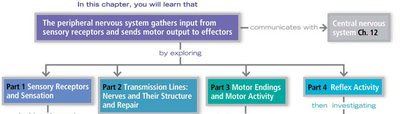
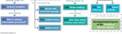
The Peripheral Nervous System (PNS) gathers input from sensory receptors and sends motor output to effectors, linking the external and internal environments to the Central Nervous System (CNS). This chapter explores sensory receptors, nerve structure and repair, motor endings, and reflex activity.
13.1 Sensory Receptors
Classification of Sensory Receptors
Sensory receptors are specialized to respond to environmental changes (stimuli). Their activation results in graded potentials that trigger nerve impulses, leading to sensation and perception in the brain. Receptors are classified by:
Stimulus Type: Mechanoreceptors (touch, pressure, vibration, stretch), Thermoreceptors (temperature), Photoreceptors (light), Chemoreceptors (chemicals), Nociceptors (pain-causing stimuli).
Location: Exteroceptors (external stimuli), Interoceptors (internal stimuli), Proprioceptors (muscle and joint stretch).
Structure: Simple (general senses) or complex (special senses).
Structural Classification of Sensory Receptors
General sensory receptors are divided into nonencapsulated (free nerve endings) and encapsulated dendritic endings. These structures determine their function and location in the body.
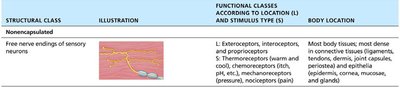
Nonencapsulated (Free) Nerve Endings
Abundant in epithelia and connective tissues.
Respond to temperature, pain, and light touch.
Include tactile (Merkel) discs and hair follicle receptors.
Encapsulated Dendritic Endings
Mechanoreceptors encased in connective tissue capsules.
Include tactile (Meissner's) corpuscles, lamellar (Pacinian) corpuscles, bulbous (Ruffini) endings, muscle spindles, tendon organs, and joint kinesthetic receptors.
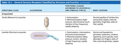
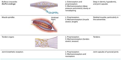
Table: General Sensory Receptors Classified by Structure and Function
Structural Class | Illustration | Functional Classes (Location & Stimulus Type) | Body Location |
|---|---|---|---|
Free nerve endings of sensory neurons | See image_3 | Exteroceptors, interoceptors, proprioceptors; thermoreceptors, chemoreceptors, mechanoreceptors, nociceptors | Most body tissues; dense in connective tissues and epithelia |
Tactile (Meissner's) corpuscles | See image_5 | Exteroceptors; mechanoreceptors (light pressure, touch, vibration) | Dermal papillae of hairless skin (fingertips, soles, eyelids) |
Lamellar (Pacinian) corpuscles | See image_5 | Exteroceptors, interoceptors, proprioceptors; mechanoreceptors (deep pressure, vibration) | Dermis, hypodermis, periostea, tendons, ligaments, joint capsules |
Bulbous corpuscles (Ruffini endings) | See image_6 | Exteroceptors, proprioceptors; mechanoreceptors (deep pressure, stretch) | Deep dermis, hypodermis, joint capsules |
Muscle spindles | See image_6 | Proprioceptors; mechanoreceptors (muscle stretch, length) | Skeletal muscles, especially extremities |
Tendon organs | See image_6 | Proprioceptors; mechanoreceptors (tendon stretch, tension) | Tendons |
Joint kinesthetic receptors | See image_6 | Proprioceptors; mechanoreceptors, nociceptors | Joint capsules of synovial joints |
13.2 Sensory Processing
Levels of Neural Integration in Sensory Systems
The somatosensory system processes sensory input at three levels:
Receptor Level: Sensory receptors generate signals when stimuli match their specificity and are within their receptive field. Transduction converts stimulus energy into graded potentials (generator or receptor potentials).
Circuit Level: Sensory impulses are conducted by three-neuron pathways to the appropriate cortical regions. First-order neurons relay from receptors to the CNS, second-order neurons transmit to the thalamus, and third-order neurons reach the somatosensory cortex.
Perceptual Level: Interpretation of sensory input occurs in the sensory cortex, including perceptual detection, magnitude estimation, spatial discrimination, feature abstraction, quality discrimination, and pattern recognition.
Adaptation of Sensory Receptors
Adaptation is a change in sensitivity in the presence of a constant stimulus. Fast-adapting receptors (e.g., pressure, touch, smell) send signals at the beginning or end of a stimulus, while slow-adapting receptors (e.g., nociceptors, most proprioceptors) adapt slowly or not at all.
Perception of Pain
Pain Mechanisms and Tolerance
Pain warns of actual or impending tissue damage. Stimuli include extreme pressure, temperature, and chemicals. Pain impulses travel on fibers releasing neurotransmitters like glutamate and substance P. Endogenous opioids (e.g., endorphins) can block some pain impulses. Pain tolerance varies among individuals and is influenced by genetics.
Visceral and Referred Pain
Visceral pain arises from internal organ receptors and is felt as vague aching or burning. Referred pain occurs when pain from one region is perceived as coming from another, due to shared nerve pathways (e.g., left arm pain during a heart attack).
13.3 Nerves
Structure of a Nerve
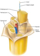
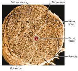
A nerve is a cordlike organ in the PNS, consisting of bundles of myelinated and nonmyelinated axons enclosed by connective tissue. The coverings include:
Endoneurium: Encloses individual axons and their myelin sheaths.
Perineurium: Bundles fibers into fascicles.
Epineurium: Surrounds all fascicles to form the nerve.
Classification of Nerves
Mixed nerves: Contain both sensory and motor fibers; impulses travel to and from CNS.
Sensory (afferent) nerves: Carry impulses toward CNS.
Motor (efferent) nerves: Carry impulses away from CNS.
Most nerves are mixed, containing somatic and autonomic fibers.
Ganglia
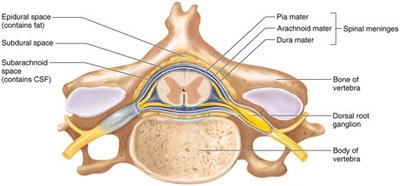
Ganglia are clusters of neuron cell bodies associated with nerves in the PNS. Sensory ganglia (dorsal root ganglia) contain cell bodies of sensory neurons, while autonomic ganglia contain motor neurons.
Regeneration of Nerve Fibers
CNS vs. PNS Regeneration
Mature neurons are amitotic. Peripheral axons may regenerate if the soma is intact, but CNS axons rarely regenerate due to inhibitory proteins and scar tissue formation by astrocytes.
PNS Axon Regeneration Process
Axon fragments and myelin sheaths distal to injury degenerate (Wallerian degeneration).
Macrophages clean up debris; Schwann cells divide.
Axon filaments grow through a regeneration tube formed by Schwann cells.
Axon regenerates and new myelin sheath forms.
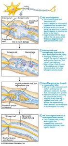
Summary Table: Steps of PNS Nerve Regeneration
Step | Description |
|---|---|
1 | Axon and myelin sheath distal to injury degenerate (Wallerian degeneration). |
2 | Schwann cells and macrophages clean up debris and stimulate Schwann cell division. |
3 | Axon filaments grow through regeneration tube formed by Schwann cells. |
4 | Axon regenerates and new myelin sheath forms. |




Key Learning Outcomes
Classify general sensory receptors by stimulus detected, body location, and structure.
Outline the events that lead to sensation and perception.
Describe receptor and generator potentials and sensory adaptation.
Describe the general structure of a nerve and the process of nerve regeneration.
Define ganglion and indicate the general body location of ganglia.
