 Back
BackPeripheral Nervous System: Structure, Function, and Integration
Study Guide - Smart Notes
 Tailored notes based on your materials, expanded with key definitions, examples, and context.
Peripheral Nervous System (PNS)
Overview
The Peripheral Nervous System (PNS) consists of all neural structures outside the brain and spinal cord. It connects the central nervous system (CNS) to limbs and organs, facilitating communication between the body and the CNS.
Components: Sensory receptors, peripheral nerves, ganglia, and motor endings
Main Functions: Sensory input, integration, and motor output
Sensory Receptors
Definition and Function
Sensory receptors are specialized structures that detect changes in the environment (stimuli) and convert them into nerve impulses. They are classified by the type of stimulus detected, location, and structural complexity.
Transduction: Conversion of stimulus energy into graded potentials, which may trigger action potentials if threshold is reached.
Classification by Stimulus Detected
Mechanoreceptors: Respond to mechanical force (touch, pressure, vibration, stretch)
Thermoreceptors: Detect changes in temperature
Photoreceptors: Respond to light (e.g., in the retina)
Chemoreceptors: Detect chemicals (e.g., taste, smell, blood chemistry)
Nociceptors: Detect pain, typically from damaging stimuli
Classification by Location
Exteroceptors: Respond to stimuli outside the body (e.g., skin, special senses)
Interoceptors (Visceroceptors): Respond to stimuli within the body (e.g., internal organs)
Proprioceptors: Detect stretch in muscles, tendons, joints, and ligaments
Classification by Structure
Free Nerve Endings (Nonencapsulated): Detect pain, temperature, and some pressure
Encapsulated Nerve Endings: Dendrites enclosed in connective tissue capsules, generally mechanoreceptors
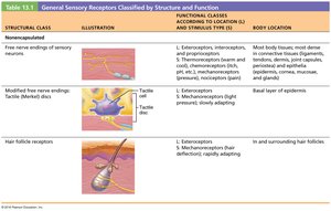
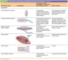
Encapsulated Receptors
Tactile (Meissner's) Corpuscles: Light touch, located in dermal papillae
Lamellar (Pacinian) Corpuscles: Deep pressure and vibration, found in dermis and subcutaneous tissue
Bulbous Corpuscles (Ruffini endings): Deep, continuous pressure, found in dermis, subcutaneous tissue, and joint capsules
Muscle Spindles: Detect muscle stretch, located in skeletal muscle perimysium
Tendon Organs: Detect tendon stretch, found in tendons
Joint Kinesthetic Receptors: Monitor joint position and movement, found in articular capsules
Sensory Integration
Levels of Sensory Integration
Sensory information is processed at three main levels:
Receptor Level: Sensory receptors transduce stimuli into graded potentials
Circuit Level: Impulses are relayed to appropriate CNS regions via ascending pathways
Perception Level: Sensory input is interpreted in the cerebral cortex
Receptor Level
Transduction: Conversion of stimulus energy to graded potential energy
Threshold: Action potential generated if threshold is reached
Frequency Coding: Frequency of nerve impulses encodes stimulus intensity
Circuit Level
First-order neurons: From receptor to spinal cord/brainstem
Second-order neurons: To cerebellum or thalamus
Third-order neurons: From thalamus to somatosensory cortex
Perception Level
Perceptual Detection: Awareness that a stimulus has occurred
Magnitude Estimation: Ability to detect intensity
Spatial Discrimination: Identifying site or pattern of stimulation
Feature Abstraction: Identifying more complex aspects of sensation
Quality Discrimination: Differentiating submodalities (e.g., sweet vs. sour)
Pattern Recognition: Recognizing familiar or significant patterns
Nerves and Nerve Structure
Connective Tissue Coverings
Endoneurium: Surrounds individual nerve fibers
Perineurium: Surrounds fascicles (bundles of fibers)
Epineurium: Surrounds the entire nerve
Types of Nerves
Sensory (Afferent) Nerves: Carry impulses to CNS
Motor (Efferent) Nerves: Carry impulses from CNS
Mixed Nerves: Contain both sensory and motor fibers (most common)
Anatomical Classes: Cranial nerves and spinal nerves
Regeneration of Nerves
PNS Axons: Can regenerate if cell body is intact and Schwann cells are functional
CNS Axons: Generally do not regenerate due to inhibitory factors and lack of supportive environment
Cranial Nerves
Overview
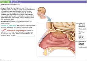
Cranial nerves emerge from the brain and primarily serve the head and neck. There are 12 pairs, each with specific sensory, motor, or mixed functions.
I – Olfactory: Sensory (smell)
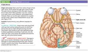
II – Optic: Sensory (vision)
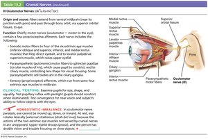
III – Oculomotor: Motor (eye movement)
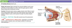
IV – Trochlear: Motor (eye movement)
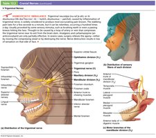
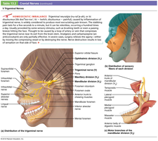
V – Trigeminal: Mixed (facial sensation, mastication)
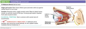
VI – Abducens: Motor (eye movement)
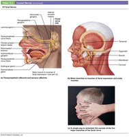
VII – Facial: Mixed (facial expression, taste)
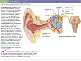
VIII – Vestibulocochlear: Sensory (hearing, balance)
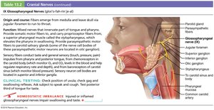
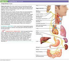
IX – Glossopharyngeal: Mixed (taste, swallowing)
X – Vagus: Mixed (viscera, heart, lungs)
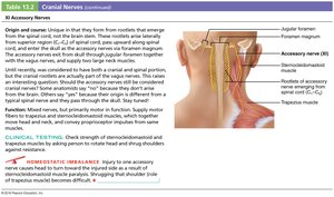
XI – Accessory: Motor (neck muscles)
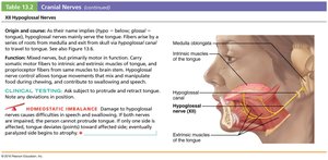
XII – Hypoglossal: Motor (tongue movement)
Spinal Nerves and Plexuses
Spinal Nerves
31 pairs: C1–C8 (cervical), T1–T12 (thoracic), L1–L5 (lumbar), S1–S5 (sacral), C0 (coccygeal)
Ventral roots: Motor (efferent) fibers
Dorsal roots: Sensory (afferent) fibers
Plexus: Network of diverging and converging nerve fibers
Plexuses
Cervical Plexus (C1–C4): Sensory from skin, motor to diaphragm (phrenic nerve)
Brachial Plexus (C5–T1): Axillary, musculocutaneous, median, ulnar, and radial nerves supply upper limb
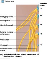
Lumbar Plexus (L1–L4): Femoral and obturator nerves supply anterior and medial thigh
Sacral Plexus (L4–S4): Pudendal and sciatic nerves supply lower limb and genitals
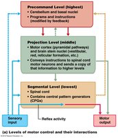
Motor Integration
Levels of Motor Control
Segmental Level: Spinal cord circuits generate basic movements and reflexes
Projection Level: Upper motor neurons in cortex and brainstem send commands to spinal cord
Precommand Level: Cerebellum and basal nuclei plan and coordinate complex movements
Reflexes and Reflex Arcs
Types of Reflexes
Intrinsic Reflex: Rapid, predictable, unlearned response to a stimulus
Acquired Reflex: Learned through practice or repetition
Reflex Arc Components
Receptor: Site of stimulus
Sensory Neuron: Transmits afferent impulses to CNS
Integration Center: Synapse(s) in CNS
Motor Neuron: Conducts efferent impulses to effector
Effector: Muscle or gland that responds
