 Back
BackPeripheral Nervous System: Structure, Function, and Clinical Relevance
Study Guide - Smart Notes
 Tailored notes based on your materials, expanded with key definitions, examples, and context.
The Peripheral Nervous System (PNS)
Overview and Structural Organization
The Peripheral Nervous System (PNS) serves as the communication link between the central nervous system (CNS) and the rest of the body. It consists of all neural structures outside the brain and spinal cord and is divided into sensory (afferent) and motor (efferent) divisions. - Sensory (afferent) division: Transmits sensory information from receptors to the CNS. - Motor (efferent) division: Transmits motor commands from the CNS to effectors (muscles and glands). - The motor division is further subdivided into the somatic nervous system (controls voluntary movements) and the autonomic nervous system (controls involuntary functions), which includes the sympathetic and parasympathetic divisions. 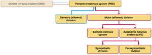
Clinical Importance
Understanding the PNS is crucial for recognizing and treating nerve damage, as it is involved in sensory perception, motor control, and reflexes.
Part 1—Sensory Receptors and Sensation
13.1 Sensory Receptors: Classification and Function
Sensory receptors are specialized cells or structures that respond to environmental changes (stimuli). Their activation generates graded potentials, which may trigger nerve impulses. Sensation (awareness of stimulus) and perception (interpretation of stimulus) occur in the brain. Classification of Sensory Receptors: 1. By Stimulus Type: - Mechanoreceptors: Respond to touch, pressure, vibration, and stretch. - Thermoreceptors: Sensitive to temperature changes. - Photoreceptors: Respond to light (e.g., retina). - Chemoreceptors: Respond to chemicals (e.g., smell, taste, blood chemistry). - Nociceptors: Sensitive to pain-causing stimuli (e.g., extreme heat/cold, pressure, chemicals). 2. By Location: - Exteroceptors: Detect external stimuli (skin, special sense organs). - Interoceptors (visceroceptors): Detect internal stimuli (viscera, blood vessels). - Proprioceptors: Detect stretch in muscles, tendons, joints, and inform the brain of movement. 3. By Structural Complexity: - Simple receptors: Modified dendritic endings of sensory neurons; monitor general sensory information. - Complex receptors: Specialized sense organs for vision, hearing, equilibrium, smell, and taste.
Structural Classification of General Sensory Receptors
Nonencapsulated (Free) Nerve Endings: - Abundant in epithelia and connective tissues. - Mostly nonmyelinated, small-diameter fibers. - Respond to temperature, pain, and light touch. - Examples: Thermoreceptors, nociceptors, tactile (Merkel) discs, hair follicle receptors. 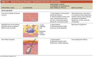 Encapsulated Dendritic Endings: - Mechanoreceptors with terminal endings encased in connective tissue. - Examples: Tactile (Meissner’s) corpuscles (discriminative touch), Lamellar (Pacinian) corpuscles (deep pressure/vibration), Bulbous (Ruffini) endings (continuous pressure), muscle spindles, tendon organs, joint kinesthetic receptors.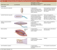
Capsaicin and Nociceptors
- Capsaicin: Chemical in hot peppers that opens ligand-gated ion channels in nociceptors, causing pain perception. - Repeated application desensitizes nociceptors, used in topical creams for neuropathic pain.
13.2 Sensory Processing
Somatosensory System: Levels of Neural Integration
The somatosensory system processes sensory input from exteroceptors, proprioceptors, and interoceptors. Sensory information is relayed toward the brain and processed at three levels: 1. Receptor level: Sensory receptors detect stimuli and generate signals. 2. Circuit level: Ascending pathways transmit signals to the brain. 3. Perceptual level: Sensory input is interpreted in cortical sensory areas. 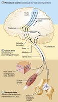
Processing at the Receptor Level
- Stimulus must match receptor specificity and be within the receptive field. - Transduction converts stimulus energy into graded potentials. - Graded potentials must reach threshold to trigger action potentials. - Adaptation: Change in sensitivity to constant stimulus. - Phasic receptors: Fast-adapting (pressure, touch, smell). - Tonic receptors: Slow-adapting or non-adapting (nociceptors, proprioceptors).
Processing at the Circuit and Perceptual Levels
- Circuit level: Three-neuron pathways conduct impulses to the cortex. - Perceptual level: Interpretation depends on the location of target neurons. - Aspects include perceptual detection, magnitude estimation, spatial discrimination, feature abstraction, quality discrimination, and pattern recognition. 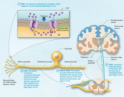
Perception of Pain
- Pain warns of tissue damage and triggers protective actions. - Pain tolerance varies among individuals; genes influence tolerance and response to medications. - Visceral pain: Originates from internal organs, often perceived as referred pain (e.g., left arm pain during heart attack). 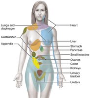
Part 2—Transmission Lines: Nerves and Their Structure and Repair
13.3 Nerves: Structure and Classification
Nerves are cordlike organs composed of bundles of myelinated and nonmyelinated axons, enclosed by connective tissue. - Connective tissue coverings: - Endoneurium: Surrounds individual axons. - Perineurium: Bundles axons into fascicles. - Epineurium: Encloses all fascicles to form the nerve. 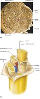
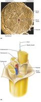 - Most nerves are mixed (contain both sensory and motor fibers). - Ganglia: Collections of neuron cell bodies in the PNS.
Regeneration of Nerve Fibers
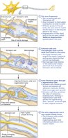
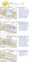
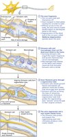
- CNS axons: Limited regeneration due to inhibitory proteins and scar tissue. - PNS axons: Can regenerate if the soma is intact. - Steps: Wallerian degeneration, debris removal by macrophages, neurolemmocyte division, axon regrowth, and new myelin formation. 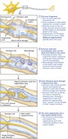
Part 3—Motor Endings and Motor Activity
13.6 Peripheral Motor Endings
Motor endings are PNS elements that activate effectors (muscles and glands) by releasing neurotransmitters. - Neuromuscular junction: Site where motor neurons communicate with skeletal muscle fibers. 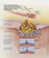
Part 4—Reflex Activity
13.8 Reflex Arc
Reflexes are rapid, involuntary responses to stimuli, classified as somatic (skeletal muscle) or autonomic (visceral effectors). Components of a Reflex Arc: 1. Receptor 2. Sensory neuron 3. Integration center 4. Motor neuron 5. Effector 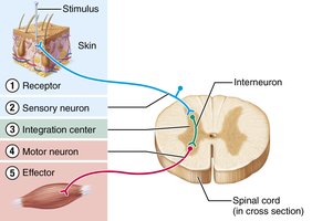
13.9 Somatic Spinal Reflexes
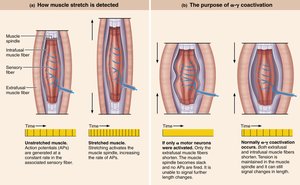
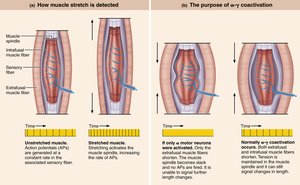
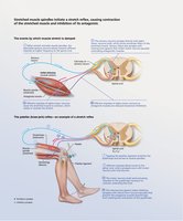
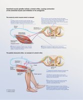
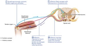
- Stretch reflex: Maintains muscle tone and posture; example is the knee-jerk reflex. - Tendon reflex: Prevents muscle damage from excessive stretch. - Flexor (withdrawal) reflex: Protects from injury by withdrawing from painful stimuli. - Crossed-extensor reflex: Maintains balance during withdrawal. 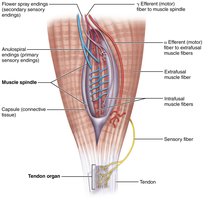
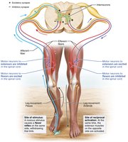
Superficial Reflexes
- Plantar reflex: Tests spinal cord integrity; abnormal response is Babinski’s sign. - Abdominal reflex: Tests cord integrity from T8 to T12. 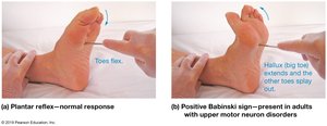
Summary Table: Major Spinal Nerve Plexuses
Dermatomes: Areas of skin innervated by specific spinal nerves; useful for assessing nerve damage. 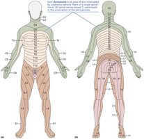
Developmental Aspects of the PNS
- Spinal nerves branch from the developing spinal cord and neural crest cells. - With age, sensory receptors atrophy, muscle tone decreases, and reflexes slow. Peripheral nerves remain viable throughout life unless subjected to trauma.
