 Back
BackPhysiology of the Respiratory System: Structured Study Notes
Study Guide - Smart Notes
 Tailored notes based on your materials, expanded with key definitions, examples, and context.
Function and Overview of the Respiratory System
General Function
The primary function of the respiratory system is to facilitate the exchange of gases, providing oxygen to tissues and cells while removing carbon dioxide from the body. This process is essential for cellular metabolism and maintaining homeostasis.
Oxygen is required for cellular respiration and energy production.
Carbon dioxide is a waste product that must be expelled to prevent acidosis.
Respiratory Physiology: Key Processes
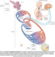
Respiratory physiology encompasses several coordinated processes:
External respiration: Exchange of gases between air in the lungs and blood.
Pulmonary ventilation: Movement of air into and out of the lungs (breathing).
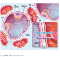
Pulmonary gas exchange: Transfer of gases across the alveolar membrane.
Transport of gases: Movement of oxygen and carbon dioxide in the blood.
Internal respiration: Exchange of gases between blood and tissues.
Cellular respiration: Utilization of oxygen by cells to produce ATP from glucose.
Regulation of respiration: Neural and chemical control of breathing rate and depth.
Anatomy of the Respiratory System
Pathway of Air
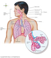
Air travels through a series of anatomical structures before reaching the alveoli, where gas exchange occurs.
Nasal cavity → Pharynx → Larynx → Trachea → Bronchi → Bronchioles → Alveoli
The Nose and Nasal Cavity
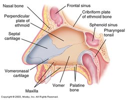
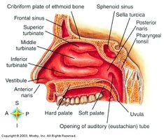
The nose and nasal cavity filter, warm, and humidify incoming air. The nasal cavity is lined with mucosa and contains structures that increase surface area for air processing.
Nasal bone, septal cartilage, vomer, maxilla, palatine bone: Structural components.
Cribriform plate of ethmoid bone: Allows passage of olfactory nerves.
Turbinates (superior, middle, inferior): Increase surface area and turbulence.
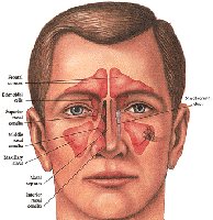
Paranasal Sinuses
Paranasal sinuses are air-filled spaces that drain into the nasal cavity. They lighten the skull and contribute to voice resonance.
Frontal, maxillary, ethmoid, sphenoid sinuses: Major sinuses.
Pharynx
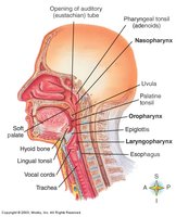
The pharynx is a muscular tube common to both the respiratory and digestive systems. It is divided into three regions:
Nasopharynx: Posterior to nasal cavity.
Oropharynx: Posterior to oral cavity.
Laryngopharynx: Posterior to larynx.
Larynx
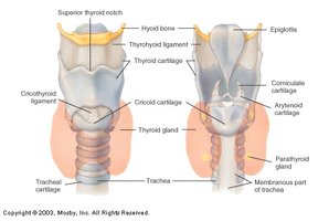
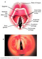
The larynx houses the vocal cords and is involved in sound production and protecting the airway during swallowing.
Thyroid cartilage, cricoid cartilage, epiglottis: Major structural components.
Vocal cords: Produce sound.
Trachea
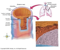
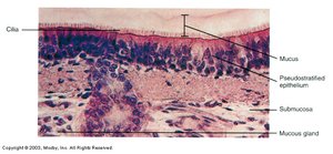
The trachea is a tube supported by cartilaginous rings, lined with ciliated pseudostratified epithelium that helps trap and move particles out of the airway.
Hyaline cartilage: Maintains airway patency.
Cilia and mucus: Trap and remove debris.
Bronchial Tubes and Lungs
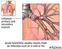
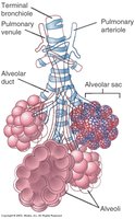
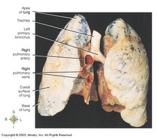
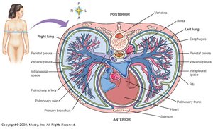
The bronchial tree branches into smaller tubes, ending in alveoli where gas exchange occurs. The lungs are divided into lobes and are surrounded by pleura.
Primary, secondary, tertiary bronchi: Progressive branching.
Alveoli: Site of gas exchange.
Pulmonary Ventilation (Breathing)
Respiratory Cycle
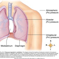
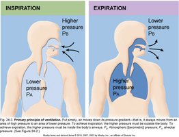
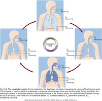
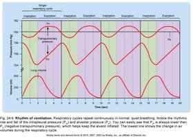
Pulmonary ventilation consists of inspiration and expiration, driven by pressure gradients between the alveoli and the atmosphere.
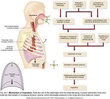
Inspiration: Air moves into lungs when alveolar pressure is lower than atmospheric pressure.
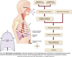
Expiration: Air moves out when alveolar pressure is higher than atmospheric pressure.
Mechanism of Pulmonary Ventilation
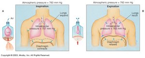
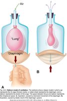
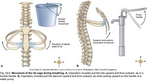
Pressure gradients are established by changes in thoracic cavity size, produced by muscle contraction and relaxation. Boyle’s law governs the relationship between volume and pressure:
Boyle’s law: (Pressure is inversely proportional to volume at constant temperature)
Diaphragm contraction: Increases thoracic volume, decreases intrapleural and alveolar pressure, causing inspiration.
Expiration: Passive process; relaxation of inspiratory muscles decreases thoracic volume, increases pressure, causing air to exit.
Compliance: Ability of lung tissues to stretch.
Elastic recoil: Tendency of lungs to return to original size after stretching.
Pulmonary Volumes and Capacities
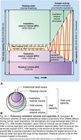
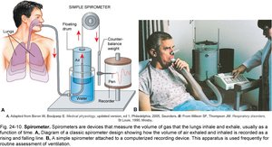
Normal gas exchange depends on adequate volumes of air moving in and out of the lungs. These volumes are measured using a spirometer.
Tidal volume (TV): Air exhaled after normal inspiration (~500 mL).
Expiratory reserve volume (ERV): Maximum air exhaled after normal expiration (1.0–1.2 L).
Inspiratory reserve volume (IRV): Maximum air inhaled after normal inspiration (3.3 L).
Residual volume: Air remaining after maximal exhalation (1.2 L).
Pulmonary Capacities
Vital capacity (VC):
Functional residual capacity (FRC): Air at end of normal respiration.
Total lung capacity (TLC): Sum of all four lung volumes.
Alveolar ventilation: Volume of inspired air reaching alveoli.
Dead Space
Anatomical dead space: Air in passageways not involved in gas exchange.
Physiological dead space: Anatomical dead space plus nonfunctioning alveoli.
Pulmonary Air Flow
Total minute volume: Volume moved per minute.
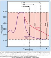
Forced expiratory volume (FEV): Volume expired per second during forced expiration.
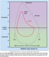
Flow-volume loop: Graphical representation of inspiratory and expiratory flow.
Pulmonary Gas Exchange
Partial Pressure and Dalton’s Law
Gas exchange is driven by partial pressure gradients. Dalton’s law states:
Law of partial pressures: (Total pressure is the sum of partial pressures of individual gases)
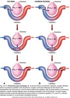
Arterial blood PO2 and PCO2 equal alveolar PO2 and PCO2.
Exchange of Gases in the Lungs
Oxygen diffuses from alveolar air to blood, and carbon dioxide diffuses from blood to alveolar air. Four factors affect oxygen diffusion:
Oxygen pressure gradient
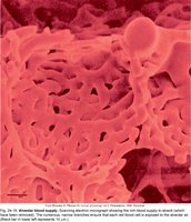
Functional surface area of respiratory membrane
Respiratory minute volume
Alveolar ventilation
Transport of Gases by the Blood
Oxygen Transport
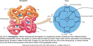
Oxygen is transported dissolved in plasma and bound to hemoglobin (Hb). Hemoglobin consists of four polypeptide chains, each with an iron-containing heme group.
Oxygen binds to iron in heme groups.
Hb increases oxygen-carrying capacity of blood.
Carbon Dioxide Transport
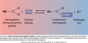
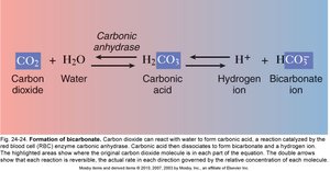
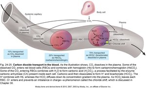
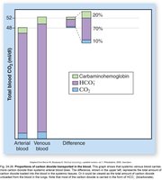
Carbon dioxide is transported in three forms:
Dissolved in plasma (10%)
Bound to hemoglobin as carbaminohemoglobin (20%)
As bicarbonate ions in plasma (70%)
Systemic Gas Exchange
Exchange in Tissues
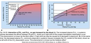
Oxygen diffuses from arterial blood to tissues, while carbon dioxide diffuses from tissues to blood. The Bohr and Haldane effects describe how these exchanges are influenced by partial pressures:
Bohr effect: Increased PCO2 decreases affinity between oxygen and Hb.
Haldane effect: Increased CO2 loading caused by decreased PO2.
Regulation of Pulmonary Function
Respiratory Control Centers
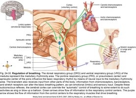
Breathing is regulated by centers in the brainstem:
Medullary rhythmicity center: Generates basic rhythm.
Inspiratory center: Stimulates inspiration.
Expiratory center: Stimulates expiration.
Apneustic center (pons): Increases length and depth of inspiration.
Pneumotaxic center (pons): Inhibits apneustic and inspiratory centers to prevent overinflation.
Factors Influencing Breathing
Breathing is influenced by chemical and neural feedback:
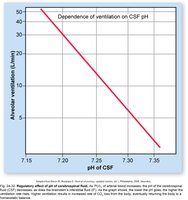
Changes in PO2, PCO2, and pH affect the medullary rhythmicity area.
PCO2 acts on central chemoreceptors; increased PCO2 leads to faster breathing.
Decreased blood pH stimulates peripheral and central chemoreceptors.
Arterial blood pressure and reflexes (Hering-Breuer) regulate depth and volume.
Cerebral cortex can voluntarily alter breathing rate and strength.
The Big Picture: Respiratory Physiology and the Whole Body
Integration with Other Systems
The respiratory system works closely with the cardiovascular, nervous, skeletal, and immune systems to maintain homeostasis:
Blood gases are transported by the cardiovascular system.
Nervous system regulates ventilation in response to internal changes.
Skeletal muscles and bones facilitate expansion and recoil of the thorax.
Immune system protects against respiratory pathogens.
Summary Table: Pulmonary Volumes and Capacities
Volume/Capacity | Definition | Normal Value |
|---|---|---|
Tidal Volume (TV) | Air exhaled after normal inspiration | ~500 mL |
Expiratory Reserve Volume (ERV) | Max air exhaled after normal expiration | 1.0–1.2 L |
Inspiratory Reserve Volume (IRV) | Max air inhaled after normal inspiration | 3.3 L |
Residual Volume | Air remaining after maximal exhalation | 1.2 L |
Vital Capacity (VC) | IRV + TV + ERV | Varies |
Total Lung Capacity (TLC) | Sum of all volumes | Varies |
Summary Table: Gas Transport Mechanisms
Gas | Transport Mechanism | Proportion |
|---|---|---|
Oxygen | Bound to hemoglobin | ~98% |
Oxygen | Dissolved in plasma | ~2% |
Carbon Dioxide | Dissolved in plasma | 10% |
Carbon Dioxide | Bound to hemoglobin (carbaminohemoglobin) | 20% |
Carbon Dioxide | As bicarbonate ions | 70% |
