 Back
BackRespiratory and Digestive System Anatomy: Lab Review Study Notes
Study Guide - Smart Notes
 Tailored notes based on your materials, expanded with key definitions, examples, and context.
Respiratory System Anatomy
Nasal and Oral Cavities
The nasal cavity and oral cavity are the initial entry points for air into the respiratory system. They play important roles in filtering, warming, and humidifying incoming air, as well as in immune defense.
Nasal conchae: Curved bony structures that increase surface area for air filtration and humidification.
Pharyngeal tonsil: Lymphatic tissue in the nasopharynx that helps trap pathogens.
Palatine tonsil: Located at the back of the oral cavity; part of the immune system.
Lingual tonsil: Found at the base of the tongue; also involved in immune defense.
Uvula: Soft tissue that helps close off the nasopharynx during swallowing.
Pharynx and Larynx
The pharynx (throat) is a muscular tube that serves both respiratory and digestive functions. The larynx (voice box) is involved in sound production and airway protection.
Nasopharynx: Upper part of the pharynx, behind the nasal cavity.
Oropharynx: Middle section, behind the oral cavity.
Laryngopharynx: Lower section, leading to the larynx and esophagus.
Epiglottis: Flap of cartilage that prevents food from entering the trachea during swallowing.
Glottis: Opening between the vocal folds.
Vocal folds: Tissue bands that vibrate to produce sound.
Thyroid and cricoid cartilage: Cartilaginous structures that support and protect the larynx.
Trachea and Bronchial Tree
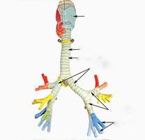
The trachea (windpipe) conducts air from the larynx to the bronchi. The bronchial tree consists of branching airways that distribute air to the lungs.
Primary (mainstem) bronchi: Right and left branches from the trachea entering each lung.
Secondary (lobar) bronchi: Branches that supply each lung lobe.
Tertiary (segmental) bronchi: Further divisions supplying bronchopulmonary segments.
Bronchioles: Smallest airways lacking cartilage; control airflow resistance (diameter <2 mm).
Alveoli: Tiny air sacs where gas exchange occurs; grouped in alveolar sacs and connected by alveolar ducts.
Lungs and Associated Structures
The lungs are paired organs responsible for gas exchange. They are divided into lobes and protected by pleural membranes.
Pleural membrane: Double-layered serous membrane enclosing each lung in a pleural cavity.
Apex and base: Apex is the superior tip; base rests on the diaphragm.
Lobes: Right lung has three lobes (superior, middle, inferior); left lung has two (superior, inferior).
Fissures: Horizontal and oblique fissures separate lung lobes.
Diaphragm: Dome-shaped muscle separating thoracic and abdominal cavities; primary muscle of respiration.
Digestive System Anatomy and Functions
Oral Cavity and Associated Structures
The oral cavity initiates mechanical and chemical digestion. Several structures contribute to this process.
Salivary glands: Three pairs (parotid, submandibular, sublingual) secrete saliva containing enzymes (e.g., amylase).
Teeth: Structures for mechanical breakdown of food; composed of enamel (hard outer layer), dentin (underlying tissue), and periodontal ligaments (anchor teeth to jaw).
Pharynx: Passageway for food and air; divided into nasopharynx, oropharynx, and laryngopharynx.
Esophagus and Stomach
The esophagus is a muscular tube transporting food to the stomach. The stomach is a muscular organ that continues digestion through churning and secretion of gastric juices.
Regions of the stomach: Cardia (entry), fundus (upper part), body (main part), pylorus (exit to duodenum).
Curvatures: Greater and lesser curvatures; the greater omentum is a fatty apron attached to the greater curvature.
Rugae: Folds in the stomach lining that allow expansion.
Functions: Mechanical mixing, protein digestion (via pepsin), and regulation of food passage to the small intestine.
Small Intestine
The small intestine is the primary site for digestion and nutrient absorption. It is divided into three regions and contains specialized structures to increase surface area.
Duodenum, jejunum, ileum: Sequential regions with distinct roles in digestion and absorption.
Circular folds (plicae) and villi: Increase surface area for absorption.
Functions: Enzymatic digestion and absorption of nutrients.
Large Intestine
The large intestine absorbs water and vitamins, and forms feces. It consists of several distinct regions.
Structures: Cecum, appendix, haustra (pouches), ascending, transverse, descending, and sigmoid colon, rectum, anus.
Functions: Water reabsorption, absorption of vitamins (e.g., vitamin K produced by bacteria), and feces formation.
Anus and Sphincters
The anus is the terminal opening of the digestive tract, controlled by two sphincters.
Internal sphincter: Involuntary smooth muscle.
External sphincter: Voluntary skeletal muscle.
Liver, Gallbladder, and Pancreas
These accessory organs play crucial roles in digestion and metabolism.
Liver: Largest internal organ; produces bile, plasma proteins, and contains phagocytic cells (Kupffer cells). Located in the right upper quadrant (RUQ).
Gallbladder: Stores and concentrates bile; releases bile into the duodenum via the common bile duct.
Pancreas: Produces digestive enzymes (delivered via the pancreatic duct) and hormones (e.g., insulin).
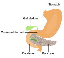
Table: Major Digestive Organs and Their Functions
Organ | Main Function(s) |
|---|---|
Oral cavity | Mechanical and chemical digestion begins |
Stomach | Protein digestion, food storage, mixing |
Small intestine | Enzymatic digestion, nutrient absorption |
Large intestine | Water/vitamin absorption, feces formation |
Liver | Bile production, metabolism, detoxification |
Gallbladder | Bile storage and concentration |
Pancreas | Digestive enzyme and hormone production |
Summary of Key Terms
Alveoli: Site of gas exchange in the lungs.
Rugae: Stomach folds for expansion.
Villi: Fingerlike projections in the small intestine for absorption.
Haustra: Pouches in the large intestine.
Kupffer cells: Liver macrophages for phagocytosis.
Additional info:
The diameter of bronchioles is typically less than 2 mm.
The right lung has three lobes, while the left has two due to the position of the heart.
Bile aids in the emulsification of fats in the small intestine.
