 Back
BackSensory Pathways and the Somatic Nervous System: Study Guide
Study Guide - Smart Notes
 Tailored notes based on your materials, expanded with key definitions, examples, and context.
Sensory Pathways and the Somatic Nervous System
Introduction
This chapter explores the organization and function of sensory pathways and the somatic nervous system (SNS). It covers the types of sensory receptors, the pathways that transmit sensory information to the central nervous system (CNS), and the motor pathways that control voluntary and involuntary movements of skeletal muscles.
General Organization of Sensory and Motor Pathways
Sensory Pathways
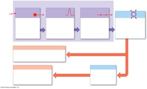
Sensory pathways are series of neurons that relay sensory information from receptors to the CNS for processing.
They utilize nerves, tracts, and nuclei (sites in the cerebral cortex).
Sensory receptors monitor specific conditions in the body or external environment and generate action potentials when stimulated.
Only about 1% of sensory information reaches the primary somatosensory cortex for conscious perception.
Nervous System Divisions
Afferent division: Somatic and visceral sensory pathways (SAME: Sensory/Afferent; Motor/Efferent).
Efferent division: Somatic motor pathways control peripheral effectors (skeletal muscles).
Sensory Receptors
Types and Functions
Sensory receptors are specialized neurons or cells monitored by sensory neurons (e.g., muscle spindle, tendon organ).
Sensation: Arriving information at the CNS.
Perception: Conscious awareness of a sensation.
General vs. Special Senses
General senses: Temperature, pain, touch, pressure, vibration, proprioception.
Special senses: Olfaction, gustation, vision, equilibrium, hearing (located in specialized sense organs).
Detection of Stimuli
Transduction: Conversion of a stimulus into an action potential by a sensory receptor.
Receptor specificity: Each receptor has a characteristic sensitivity (e.g., pressure vs. chemical).
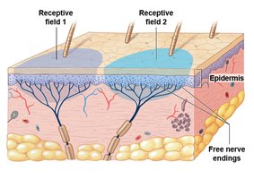
Receptive field: Area monitored by a single receptor cell; larger fields make localization more difficult.
Interpretation and Adaptation
Labeled line: Pathway from peripheral receptor to cortical neuron, carrying information about one modality.
Adaptation: Reduction in receptor sensitivity from a constant, painless stimulus.
Peripheral adaptation: Reduces information reaching the CNS.
Central adaptation: Subconscious restriction of information to the cerebral cortex.
Tonic vs. Phasic Receptors
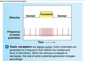
Tonic receptors: Always active, slow to adapt, e.g., pain and proprioceptors.
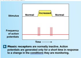
Phasic receptors: Normally inactive, fast-adapting, respond to changes in stimulus, e.g., some tactile receptors.
Classification of Sensory Receptors
By Location
Exteroceptors: External environment.
Proprioceptors: Position of skeletal muscles and joints.
Interoceptors: Visceral organs and functions.
By Nature of Stimulus
Nociceptors: Pain receptors, free nerve endings, tonic, respond to temperature extremes, mechanical damage, and chemicals.
Thermoreceptors: Temperature receptors, free nerve endings, phasic, more cold than heat receptors, found in dermis, muscles, liver, hypothalamus.
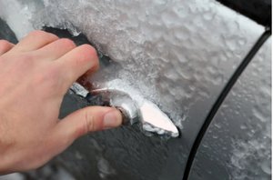
Mechanoreceptors: Respond to physical distortion (stretch, compression, twisting).
Chemoreceptors: Respond to dissolved substances, monitor pH, CO2, and O2 in blood.
Mechanoreceptor Subtypes
Tactile receptors: Touch, pressure, vibration.
Baroreceptors: Pressure changes in blood vessels and organs.
Proprioceptors: Position of joints and muscles.
Tactile Receptors in the Skin
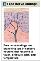
Free nerve endings: Touch, pressure, pain, temperature (tonic, small receptive fields).
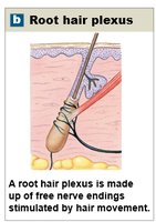
Root hair plexus: Hair movement (phasic).
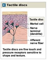
Tactile discs (Merkel discs): Fine touch, pressure, shape, texture (tonic, tiny receptive fields).
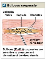
Bulbous corpuscles (Ruffini): Pressure, stretch (tonic, deep dermis).
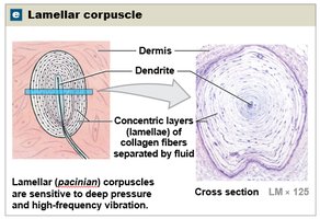
Lamellar corpuscles (Pacinian): Deep pressure, high-frequency vibration (phasic, deep dermis).
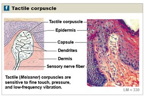
Tactile corpuscles (Meissner): Fine touch, pressure, low-frequency vibration (phasic, superficial dermis).
Proprioceptors
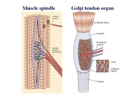
Muscle spindles: Monitor muscle length, trigger stretch reflexes.
Golgi tendon organs: Monitor tension in tendons during muscle contraction.
Joint capsule receptors: Detect pressure, tension, movement at joints.
Chemoreceptors
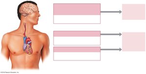
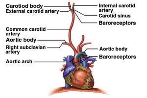
Located in carotid and aortic bodies, and medulla oblongata.
Monitor pH, CO2, and O2 in blood and cerebrospinal fluid.
Regulate respiratory and cardiovascular activity.
Somatic Sensory Pathways
Neuronal Organization
First-order neuron: Sensory neuron delivering information to the CNS (cell body in dorsal root or cranial nerve ganglion).
Second-order neuron: Interneuron in the CNS (spinal cord or brainstem).
Third-order neuron: In the thalamus, relays information to the primary somatosensory cortex (if conscious awareness is required).
Decussation: Crossing over of axons to the opposite side of the CNS.
Major Somatic Sensory Pathways
Spinothalamic pathway: Crude touch, pressure, pain, temperature.
Posterior column pathway: Fine touch, vibration, pressure, proprioception.
Spinocerebellar pathway: Proprioceptive information to the cerebellum (not consciously perceived).
Spinothalamic Pathway
First-order neurons synapse in posterior horns of spinal cord.
Second-order neurons cross to the opposite side and ascend to the thalamus.
Third-order neurons project to the primary somatosensory cortex.
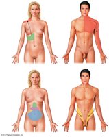
Clinical relevance: Phantom limb syndrome and referred pain (pain perceived at a location other than the site of origin).
Posterior Column Pathway
Transmits fine touch, vibration, pressure, and proprioception.
First-order neurons ascend to the medulla oblongata (gracile and cuneate fasciculi).
Second-order neurons decussate and ascend to the thalamus via the medial lemniscus.
Third-order neurons project to the primary sensory cortex.
Spinocerebellar Pathway
Conveys proprioceptive information from muscles, tendons, and joints to the cerebellum.
Does not reach conscious awareness.
Posterior tracts do not cross; anterior tracts cross twice (spinal cord and cerebellum).
Visceral Sensory Pathways
Information from interoceptors in thoracic and abdominopelvic cavities.
Ascends with spinothalamic pathway; processed in the solitary nucleus of the medulla oblongata.
Somatic Motor Pathways
Overview
Somatic Nervous System (SNS): Controls voluntary contractions of skeletal muscles.
Motor pathways always involve at least two neurons: upper motor neuron (CNS) and lower motor neuron (brainstem or spinal cord to muscle).
Basal nuclei and cerebellum provide coordination and feedback control.
Major Motor Pathways
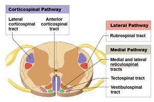
Corticospinal pathway (pyramidal system): Voluntary control over skeletal muscles; includes corticobulbar, lateral corticospinal, and anterior corticospinal tracts.
Medial pathway: Controls gross movements of trunk and proximal limbs (vestibulospinal, tectospinal, reticulospinal tracts).
Lateral pathway: Controls precise movements of distal limbs (rubrospinal tract).
Motor Homunculus
Functional map of the primary motor cortex showing the degree of fine motor control for different body regions.
Regions with more precise control (hands, face, tongue) occupy larger areas.
Basal Nuclei and Cerebellum
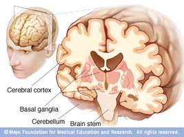
Basal nuclei: Provide background patterns of movement, adjust activities of upper motor neurons.
Cerebellum: Monitors proprioceptive, visual, and vestibular information; adjusts motor output for smooth, coordinated movements.
Summary Table: Principal Ascending (Sensory) Pathways
Pathway/Tract | Sensations | First Order | Second Order | Third Order | Final Destination | Site of Decussation |
|---|---|---|---|---|---|---|
Lateral spinothalamic | Pain and temperature | Dorsal root ganglia | Interneurons in posterior gray horns | Ventral nuclei of thalamus | Primary sensory cortex on side opposite stimulus | Axons of second-order neurons at level of entry |
Anterior spinothalamic | Crude touch and pressure | Dorsal root ganglia | Interneurons in posterior gray horns | Ventral nuclei of thalamus | Primary sensory cortex on side opposite stimulus | Axons of second-order neurons at level of entry |
Posterior column (gracile/cuneate fasciculi) | Fine touch, vibration, pressure, proprioception | Dorsal root ganglia | Nucleus gracilis/cuneatus in medulla oblongata | Ventral nuclei of thalamus | Primary sensory cortex on side opposite stimulus | Axons of second-order neurons before entering medial lemniscus |
Spinocerebellar | Proprioception | Dorsal root ganglia | Interneurons in spinal cord | Not present | Cerebellar cortex | None or twice (anterior tract) |
Additional info: Table entries inferred and summarized from textbook context.
Summary Table: Principal Descending (Motor) Pathways
Tract | Location of Upper Motor Neurons | Destination | Site of Decussation | Action |
|---|---|---|---|---|
Vestibulospinal | Vestibular nuclei at border of pons and medulla | Lower motor neurons of anterior gray horns of spinal cord | None (uncrossed) | Subconscious regulation of balance and muscle tone |
Tectospinal | Superior and inferior colliculi | Lower motor neurons of anterior gray horns of upper cervical spinal cord | Brain stem (midbrain) | Subconscious regulation of eye, head, neck, and upper limb position in response to visual and auditory stimuli |
Reticulospinal | Reticular formation (brainstem) | Lower motor neurons of anterior gray horns of spinal cord | None (uncrossed) | Subconscious regulation of reflex activity |
Rubrospinal | Red nuclei of midbrain | Lower motor neurons of anterior gray horns of cervical spinal cord | Brain stem (midbrain) | Subconscious regulation of upper limb muscle tone and movement |
Additional info: Table entries inferred and summarized from textbook context.
