 Back
BackSkeletal Muscle Contraction: Action Potentials, Neuromuscular Junction, and Excitation-Contraction Coupling
Study Guide - Smart Notes
 Tailored notes based on your materials, expanded with key definitions, examples, and context.
CH 10 PT 3 - Action Potentials in Skeletal Muscle
Stages of the Action Potential
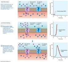
The action potential is a rapid change in membrane potential that enables communication and contraction in skeletal muscle fibers. It consists of three main stages:
Depolarization Stage: In response to a stimulus, voltage-gated sodium ion channels open, allowing sodium ions (Na+) to enter the cell. This influx makes the membrane potential less negative, reaching a peak of about +30 mV. The membrane is now less polarized, and the inside becomes positive relative to the outside.
Repolarization Stage: Sodium ion channels close, and voltage-gated potassium ion channels open. Potassium ions (K+) exit the cell, restoring the membrane potential to a more negative value, typically around −90 mV.
Propagation: The action potential is conducted throughout the sarcolemma, including the T-tubules, allowing a single stimulus to have a widespread effect.
Example: The rapid transmission of an action potential along a muscle fiber enables coordinated contraction of the entire muscle.
The Neuromuscular Junction (NMJ)
Structure and Function
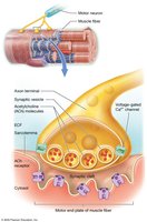
The neuromuscular junction is the synapse between a motor neuron and a skeletal muscle fiber. It is essential for transmitting the neuronal action potential to the muscle fiber, initiating contraction.
Axon Terminal (Synaptic Bulb): The swollen end of the neuron’s axon, containing synaptic vesicles filled with neurotransmitters (primarily acetylcholine (ACh)).
Synaptic Cleft: The narrow space between the axon terminal and the muscle fiber, filled with collagen fibers and extracellular gel. It contains enzymes that break down ACh.
Motor End Plate: Specialized region of the sarcolemma with many receptors for ACh, facilitating the response to neuronal signals.
Example: The release of ACh from the axon terminal triggers muscle fiber activation.
Phases of Skeletal Muscle Contraction
Excitation Phase
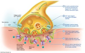
Muscle contraction begins with the excitation phase, where the sarcolemma is stimulated by ACh from a motor neuron.
An action potential arrives at the axon terminal, opening voltage-gated calcium ion channels.
Calcium ions enter the axon terminal, triggering exocytosis of synaptic vesicles.
Synaptic vesicles release ACh into the synaptic cleft.
ACh binds to ligand-gated cation channels in the motor end plate.
Cation channels open, allowing sodium ions to enter and potassium ions to exit, resulting in a net gain of positive charges.
Entry of cations depolarizes the sarcolemma locally, producing an end-plate potential.
Example: Multiple end-plate potentials are required to produce a functional contraction.
Excitation-Contraction Coupling
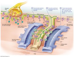
This phase transmits the excitation to the myofilaments, leading to contraction.
The end-plate potential stimulates an action potential in the sarcolemma.
The action potential is propagated down the T-tubules.
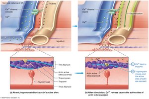
T-tubule depolarization opens calcium ion channels in the sarcoplasmic reticulum (SR), releasing calcium ions into the cytosol.
Example: Calcium ions released from the SR are essential for muscle contraction.
Contraction Phase: The Crossbridge Cycle
The contraction phase involves the sliding-filament mechanism, where myofilaments interact to produce tension.
Calcium ions bind to troponin, which has three subunits: one binds calcium, one binds actin, and one binds troponin itself.
Tropomyosin moves, exposing the active sites of actin.
Myosin heads bind to actin, forming a crossbridge and initiating the crossbridge cycle.
Example: Muscle contractions are a series of crossbridge cycles, resulting in tension and movement.
ATP Hydrolysis and Crossbridge Formation
ATP hydrolysis is critical for the crossbridge cycle:
Myosin heads bind ATP, and the ATPase enzyme catalyzes its hydrolysis to ADP and phosphate (Pi).
The energy released “cocks” the myosin head into a high-energy position, ready to bind actin.
The myosin head binds to actin at a 90° angle, forming a crossbridge.
Equation:
Example: The hydrolysis of ATP provides the energy required for muscle contraction.
Additional info: The notes above expand on the original content by providing definitions, examples, and a relevant equation for ATP hydrolysis. The images included directly reinforce the explanations of action potentials, neuromuscular junction structure, excitation phase, excitation-contraction coupling, and preparation for contraction.
