 Back
BackSkeletal Muscle Contraction, Relaxation, and Energy Sources
Study Guide - Smart Notes
 Tailored notes based on your materials, expanded with key definitions, examples, and context.
CH 10 PT 4 - Skeletal Muscle Contraction
The Crossbridge Cycle and Sliding-Filament Mechanism
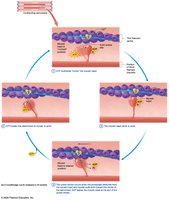
The contraction of skeletal muscle fibers is driven by the crossbridge cycle, a series of molecular events that enable the sliding of actin (thin) filaments past myosin (thick) filaments, resulting in muscle shortening and force generation.
Power Stroke: The power stroke occurs when inorganic phosphate (Pi) detaches from the myosin head, causing the myosin to pivot and pull the actin filament toward the center of the sarcomere. ADP then leaves the myosin head at the end of the power stroke.
ATP Binding and Detachment: ATP binds to the myosin head, breaking the attachment between myosin and actin. The myosin head is then recocked as ATP is hydrolyzed, allowing it to bind to the next actin subunit and repeat the cycle.
Cycle Repetition: Each myosin head undergoes this cycle 20–40 times during a single contraction, resulting in progressive shortening of the sarcomere.
Linking the Crossbridge Cycle to the Sliding-Filament Mechanism
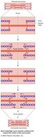
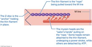
During contraction, myosin heads attach to actin and pull the thin filament toward the M line, increasing the zone of overlap and shortening the sarcomere. The process is analogous to sailors pulling a rope (thin filament) toward an anchor (Z-disc), with some hands (myosin heads) always holding and pulling while others reposition for the next pull. This coordination prevents the thin filament from sliding backward.
Muscle Relaxation
Mechanisms of Muscle Relaxation
Muscle relaxation is a multi-step process that returns the muscle fiber to its resting state after contraction:
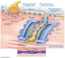
Acetylcholine Breakdown: Acetylcholinesterase (AChE) degrades acetylcholine (ACh) in the synaptic cleft, stopping stimulation of the muscle fiber.
Restoration of Membrane Potential: The sarcolemma returns to its resting membrane potential, and calcium ion channels in the sarcoplasmic reticulum (SR) close.
Calcium Reuptake: Calcium ions are actively pumped back into the SR, reducing cytosolic calcium concentration.
Troponin and Tropomyosin Reset: Troponin shifts, pulling tropomyosin back to block actin's active sites, ending contraction and allowing relaxation.
Muscle Spasms and Clinical Relevance
Continuous Activity: Calcium pumps and AChE are always active to ensure muscle fibers can relax and prepare for new contractions.
Spasm: Inability to relax leads to muscle spasm, which can be caused by dehydration, electrolyte imbalance, injury, or overload.
Rigor Mortis
Rigor mortis is the progressive stiffening of skeletal muscles after death, beginning 3–4 hours postmortem. This occurs because ATP is no longer available to fuel calcium pumps or detach myosin from actin, resulting in sustained contraction until proteins degrade (48–72 hours).

Energy Sources for Skeletal Muscle
ATP Requirement and Regeneration
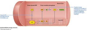
ATP is essential for maintaining ion gradients, contraction, and relaxation in muscle fibers. Since stored ATP is limited, muscle fibers regenerate ATP through three main processes:
Immediate Regeneration: Creatine phosphate (CP) donates a phosphate group to ADP, rapidly forming ATP via the enzyme creatine kinase (CK). This supplies energy for about 10 seconds of maximal activity.
Glycolytic Catabolism: Occurs in the cytosol, breaking down glucose to generate ATP anaerobically.
Oxidative Catabolism: Occurs in mitochondria, using oxygen to produce ATP from various substrates.
Creatine Supplementation: Evidence and Risks
Performance: Creatine supplementation can mildly improve performance in short, high-intensity activities but has little effect on endurance.
Risks: Excessive creatine intake can cause kidney damage and unnecessary weight gain due to water retention. Muscles have a storage limit, so excess is excreted.
Regulation: Creatine is regulated as a food, not a drug, so quality control varies and FDA approval is not required.
Summary Table: Immediate Energy Sources for Muscle Contraction
Source | Location | Duration of Supply | Key Enzyme |
|---|---|---|---|
Stored ATP | Cytosol | Few seconds | — |
Creatine Phosphate | Cytosol | ~10 seconds | Creatine Kinase |
Glycolytic Catabolism | Cytosol | 30–40 seconds | Multiple glycolytic enzymes |
Oxidative Catabolism | Mitochondria | Minutes to hours | Multiple mitochondrial enzymes |
Key Equations
ATP Hydrolysis:
Creatine Phosphate Reaction:
