 Back
BackA&P Exam 3
Study Guide - Smart Notes
 Tailored notes based on your materials, expanded with key definitions, examples, and context.
Skeletal Muscle Structure and Organization
Hierarchy of Skeletal Muscle Structure
Skeletal muscle is organized into a hierarchy of structures, each with specific roles in contraction and force generation.
Muscle: Composed of bundles of fascicles, surrounded by the epimysium.
Fascicle: Bundle of muscle fibers, surrounded by the perimysium.
Muscle Fiber (Cell): Surrounded by the endomysium; contains myofibrils.
Myofibril: Composed of repeating units called sarcomeres.
Sarcomere: The smallest contractile (functional) unit of muscle; aligned end-to-end along myofibrils.
Myofilaments: Actin (thin) and myosin (thick) filaments within sarcomeres.
Epimysium surrounds the entire muscle, perimysium surrounds each fascicle, and endomysium surrounds each muscle fiber.
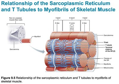
Sarcomere Structure
The sarcomere is defined as the region between two Z discs. It contains:
A band: Dark region containing thick filaments (myosin).
I band: Light region containing thin filaments (actin) only; spans two adjacent sarcomeres.
H zone: Central region of the A band with only thick filaments.
M line: Center of the H zone; holds thick filaments together.
Z disc: Boundary of each sarcomere; anchors thin filaments.
Muscle Contraction: Mechanisms and Steps
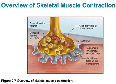
Events at the Neuromuscular Junction (NMJ)
The neuromuscular junction is the synapse between a motor neuron and a skeletal muscle fiber. It is essential for initiating muscle contraction.
Motor neuron action potential arrives at the axon terminal.
Acetylcholine (ACh) is released into the synaptic cleft.
ACh binds to receptors on the sarcolemma, causing depolarization.
Action potential is generated in the muscle fiber.
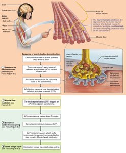
Steps of Skeletal Muscle Contraction
Four main steps are required for skeletal muscle contraction:
Events at the neuromuscular junction
Muscle fiber excitation
Excitation-contraction coupling
Cross bridge cycling
During excitation-contraction coupling, Ca2+ is released from the sarcoplasmic reticulum (SR) and binds to troponin, allowing myosin to bind to actin and initiate contraction.
Sliding Filament Model
Muscle contraction occurs as myosin heads bind to actin, pulling thin filaments toward the center of the sarcomere. This shortens the sarcomere and generates force.
Cross bridge cycle: Myosin binds to actin, performs a power stroke, detaches, and re-cocks using ATP.
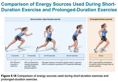
Muscle Energy Metabolism
ATP Regeneration in Muscle
Muscle fibers regenerate ATP by three main mechanisms:
Direct phosphorylation of ADP by creatine phosphate (CP): Provides energy for about 15 seconds.
Anaerobic pathway: Glycolysis and lactic acid formation; supports short, intense activity.
Aerobic pathway: Glycolysis followed by aerobic respiration in mitochondria; supports prolonged activity.
Creatine kinase is the enzyme that transfers phosphate from CP to ADP to form ATP.
Muscle Actions and Levers
Muscle Roles
Prime mover (agonist): Main muscle responsible for movement.
Antagonist: Opposes the prime mover.
Synergist: Assists the prime mover.
Lever Systems in the Body
Levers amplify force or speed of movement. There are three classes:
First-class lever: Fulcrum between effort and load (e.g., neck muscles moving the head).
Second-class lever: Load between fulcrum and effort (e.g., standing on tiptoes).
Third-class lever: Effort between fulcrum and load (most common in the body; e.g., biceps brachii flexing the forearm).
Mnemonic: "The EFL tower had an ELF but he FEL" (Effort-Fulcrum-Load order for each class).
Muscle Movements and Nerve Supply
Major Muscle Movements
Anterior muscles: Flexion
Posterior muscles: Extension
Medial muscles: Adduction
Lateral muscles: Abduction
Examples:
Chewing: Masseter and temporalis (innervated by cranial nerve V)
Breathing: Diaphragm, external and internal intercostals
Punching/pushing: Serratus anterior, pectoralis major
Special Movements
Dorsiflexion: Lifting the foot upward
Plantarflexion: Pointing the foot downward
Inversion: Turning the sole of the foot inward
Eversion: Turning the sole of the foot outward
Cranial Nerves and Muscle Innervation
Cranial Nerve V (Trigeminal): Muscles of mastication
Cranial Nerve VII (Facial): Muscles of facial expression
Cranial Nerve XI (Accessory): Sternocleidomastoid and trapezius
If these nerves are damaged, corresponding muscle functions are impaired (e.g., inability to chew, facial droop, or weak shoulder shrug).
Nervous System Overview
CNS vs PNS
Central Nervous System (CNS): Brain and spinal cord; integrates and processes information.
Peripheral Nervous System (PNS): Nerves outside the CNS; transmits signals to and from the CNS.
Localized symptoms suggest PNS issues; widespread or cognitive symptoms suggest CNS dysfunction.
Neuron Types and Locations
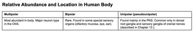
Multipolar | Bipolar | Unipolar (pseudounipolar) | |
|---|---|---|---|
Relative Abundance and Location | Most abundant in body. Major neuron type in the CNS. | Rare. Found in some special sensory organs (olfactory mucosa, eye, ear). | Found mainly in the PNS. Common only in dorsal root ganglia and sensory ganglia of cranial nerves. |
Myelin Sheath
The myelin sheath insulates axons, speeding up neural transmission. Damage to myelin (as in multiple sclerosis) slows or blocks nerve signals, causing muscle weakness, coordination problems, or sensory deficits.
Neural Transmission and Channels
Chemically gated channels: Open in response to neurotransmitters (e.g., at the NMJ).
Voltage-gated channels: Open in response to changes in membrane potential (e.g., along axons).
Somatic vs Autonomic Nervous System
Somatic: Voluntary control of skeletal muscles.
Autonomic: Involuntary control (e.g., heart, glands).
Parasympathetic: "Peace"—rest and digest.
Sympathetic: "Stress"—fight or flight.
Clinical Correlations and Disorders
Astrocytes help capture and recycle excess ACh at synapses.
Muscle weakness can result from nerve damage, myelin loss, or impaired NMJ function.
Hyperexcitability may occur if inhibitory signals are lost or if ion channel function is abnormal.
Pelvic floor dysfunction can lead to incontinence or organ prolapse; innervated by the pudendal nerve.
Rotator cuff muscles stabilize the shoulder; injury leads to weakness or limited movement.
Muscle Naming Conventions
Muscles are named based on location, shape, size, direction of fibers, number of origins, location of attachments, and action (e.g., rectus abdominis, biceps brachii).
Additional info: This guide expands on brief notes with academic context, definitions, and examples for clarity and exam preparation.
