 Back
BackSpecial Senses: Hearing and Equilibrium – Anatomy & Physiology Study Notes
Study Guide - Smart Notes
 Tailored notes based on your materials, expanded with key definitions, examples, and context.
Special Senses: Hearing and Equilibrium
Overview
The special senses of hearing and equilibrium are mediated by complex structures within the ear. This section covers the anatomy and physiology of the ear, the mechanisms of hearing and balance, and clinical tests used to assess auditory and vestibular function.
Anatomy of the Ear
Divisions of the Ear
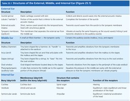
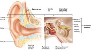
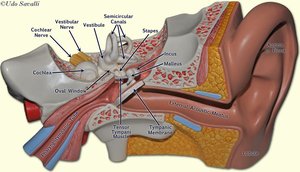
The ear is divided into three main regions: the external ear, middle ear, and internal ear. Each region contains specialized structures essential for hearing and equilibrium.
Section | Structure | Description | Function |
|---|---|---|---|
External Ear | Auricle (pinna) | Elastic cartilage covered with skin | Collects and directs sound waves into the external acoustic meatus |
External acoustic meatus | Canal lined with skin, hairs, and ceruminous glands | Transmits sound waves to the tympanic membrane | |
Tympanic membrane | Thin membrane separating external and middle ear | Vibrates in response to sound waves | |
Middle Ear | Malleus (hammer) | Bone attached to tympanic membrane | Transmits and amplifies vibrations from tympanic membrane to incus |
Incus (anvil) | Bone between malleus and stapes | Transmits and amplifies vibrations from malleus to stapes | |
Stapes (stirrup) | Bone attached to oval window | Transmits vibrations from incus to oval window of inner ear | |
Pharyngotympanic (auditory) tube | Connects middle ear to nasopharynx | Equalizes pressure in middle ear | |
Internal Ear | Cochlea | Spiral-shaped, contains cochlear duct | Hearing (contains organ of Corti) |
Vestibule | Central part, contains utricle and saccule | Static equilibrium | |
Semicircular canals | Three canals oriented in different planes | Dynamic equilibrium |
Ossicles of the Middle Ear
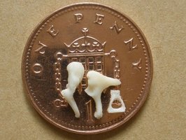
Malleus (hammer): Attached to the tympanic membrane.
Incus (anvil): Interposed between malleus and stapes.
Stapes (stirrup): Base fits into the oval window of the inner ear.
These tiny bones transmit and amplify sound vibrations from the tympanic membrane to the inner ear.
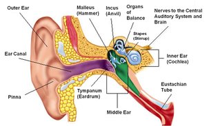
Gross Anatomy of the Ear
Understanding the spatial relationships of the ear's structures is essential for grasping their functions in hearing and balance.
Internal Ear (Labyrinth)
Major Components
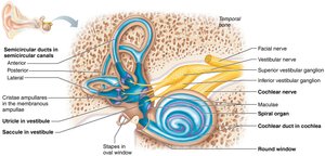
Cochlea: Responsible for hearing; contains the organ of Corti.
Vestibule: Contains utricle and saccule; involved in static equilibrium.
Semicircular Canals: Detect rotational (dynamic) movements.
Anatomy of the Cochlea
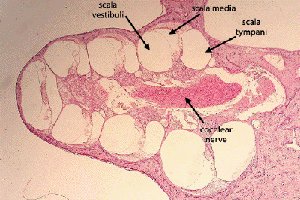
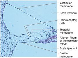
The cochlea is a spiral-shaped organ divided into three parallel ducts:
Scala vestibuli (vestibular duct): Contains perilymph, above the cochlear duct.
Scala media (cochlear duct): Contains endolymph, houses the organ of Corti.
Scala tympani (tympanic duct): Contains perilymph, below the cochlear duct.
Organ of Corti (Spiral Organ)
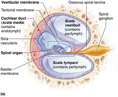
The organ of Corti is the sensory receptor for hearing, located within the cochlear duct. It consists of:
Hair cells: Sensory cells that transduce mechanical vibrations into nerve impulses.
Tectorial membrane: Overlies the hair cells and is involved in the stimulation of these cells.
Basilar membrane: Supports the organ of Corti and vibrates in response to sound.
Mechanisms of Hearing
Sound Transmission Pathway
Sound waves enter the external acoustic meatus and vibrate the tympanic membrane.
Vibrations are transmitted through the ossicles (malleus, incus, stapes) to the oval window.
Movement of the stapes at the oval window creates pressure waves in the perilymph of the scala vestibuli.
Pressure waves cause vibration of the basilar membrane, stimulating hair cells in the organ of Corti.
Hair cells generate nerve impulses that are transmitted via the cochlear branch of the vestibulocochlear nerve (CN VIII) to the brain.
Localization of Sound
Sound localization is achieved by comparing the intensity and timing of sounds reaching both ears.
The brain uses these differences to determine the direction of the sound source.
Hearing Tests
Common Clinical Tests
Weber Test: Assesses lateralization of sound to detect unilateral hearing loss.
Rinne Test: Compares air conduction to bone conduction of sound.
Audiometry: Measures hearing acuity across different frequencies.
Sound Localization Test: Determines the ability to localize sound sources.
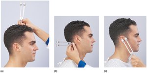
Sensorineural deafness results from damage to the inner ear or auditory nerve, while conduction deafness is due to problems in the external or middle ear. Nystagmus is an involuntary, rapid movement of the eyes, often associated with vestibular dysfunction.
Equilibrium and Balance
Types of Equilibrium
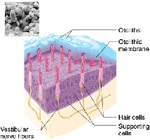
Static equilibrium: Maintains the position of the body and head when stationary; detected by the maculae in the vestibule (utricle and saccule).
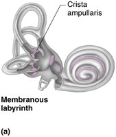
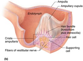
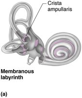
Dynamic equilibrium: Maintains balance during movement; detected by the cristae ampullares in the semicircular canals.
Semicircular Canals and Crista Ampullaris
The semicircular canals are oriented in three planes and contain the crista ampullaris, a receptor for dynamic equilibrium. Movement of endolymph within the canals bends the hair cells, generating nerve impulses.
Vestibule and Maculae
The vestibule contains the utricle and saccule, each with a macula receptor. The maculae detect linear acceleration and gravitational forces via hair cells embedded in an otolithic membrane.
Equilibrium Tests
Barany Test: Assesses the function of the semicircular canals and dynamic equilibrium.
Romberg Test: Evaluates static equilibrium and the role of vision in balance.
Summary Table: Key Structures and Functions
Structure | Location | Function |
|---|---|---|
Organ of Corti | Cochlear duct | Hearing (sound transduction) |
Crista ampullaris | Ampulla of semicircular canals | Dynamic equilibrium (rotational movement) |
Macula | Utricle and saccule of vestibule | Static equilibrium (linear acceleration, gravity) |
Key Terms and Definitions
Sensorineural deafness: Hearing loss due to damage of the inner ear or auditory nerve.
Conduction deafness: Hearing loss due to problems in the external or middle ear.
Nystagmus: Involuntary, rapid eye movements, often related to vestibular dysfunction.
Sample Questions for Review
Is the Romberg test used to assess static equilibrium or dynamic equilibrium?
What is the purpose of the Weber test?
Name the ossicle which is attached to the labyrinth.
How is the Barany test performed?
Name the fluid inside the cochlear duct (scala media). Answer: Endolymph
Name the receptor for dynamic equilibrium. Answer: Crista ampullaris
