 Back
BackSpinal Cord and Spinal Nerves: Structure, Function, and Organization
Study Guide - Smart Notes
 Tailored notes based on your materials, expanded with key definitions, examples, and context.
Spinal Cord & Spinal Nerves
Overview and Objectives
The spinal cord is a critical component of the central nervous system, serving as the main pathway for information connecting the brain and peripheral nervous system. This section covers the anatomy, protective coverings, and organization of the spinal cord and spinal nerves, including their functional significance and clinical relevance.
Anatomy of the Spinal Cord
Gross Structure and Major Regions
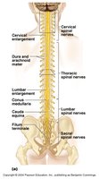
Location: The spinal cord is enclosed within the vertebral canal, extending from the foramen magnum to the level of L1 or L2 vertebra in adults.
Spinal Nerves: There are 31 pairs of spinal nerves emerging from the spinal cord, each corresponding to a segment of the vertebral column.
Enlargements: The spinal cord exhibits two major enlargements:
Cervical enlargement: Supplies nerves to the upper limbs.
Lumbar enlargement: Supplies nerves to the lower limbs.
Conus Medullaris: The tapered, conical end of the spinal cord, located below the lumbar enlargement.
Cauda Equina: A bundle of spinal nerve roots extending below the conus medullaris, resembling a horse’s tail.
Filum Terminale: A thin thread of pia mater extending from the conus medullaris to the coccyx, anchoring the spinal cord.
Protective Coverings: The Meninges
Structure and Function
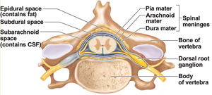
The spinal cord is protected by three connective tissue membranes known as the meninges:
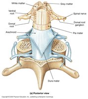
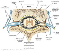
Dura Mater: The tough, outermost layer. The epidural space between the dura mater and vertebra contains fat and blood vessels and is the site for epidural anesthesia.
Arachnoid Mater: The middle, web-like layer. The subdural space lies between the dura and arachnoid, while the subarachnoid space (between arachnoid and pia) is filled with cerebrospinal fluid (CSF).
Pia Mater: The delicate, innermost layer that clings tightly to the spinal cord. The filum terminale is a specialization of the pia mater.
Internal Anatomy of the Spinal Cord
White Matter and Gray Matter
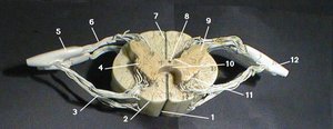
White Matter: The outer region, composed of myelinated axons organized into columns (funiculi). These columns are further divided into sensory (ascending) and motor (descending) tracts.
Gray Matter: The inner, butterfly-shaped region containing neuron cell bodies, unmyelinated fibers, and neuroglia. It is divided into:
Posterior (Dorsal) Horns: Contain interneurons and receive somatic and visceral sensory input from the dorsal root ganglia.
Anterior (Ventral) Horns: Contain cell bodies of somatic motor neurons whose axons exit via the ventral root.
Lateral Horns: Present only in thoracic and lumbar regions; contain visceral motor neurons.
Gray Commissure: Connects the two sides of gray matter and surrounds the central canal, which contains CSF.
Spinal Nerves: Structure and Organization
Formation and Branching
Roots: Each spinal nerve forms from the fusion of a dorsal (sensory) root and a ventral (motor) root.
Spinal Nerve Proper: The short segment where dorsal and ventral roots merge; all spinal nerves are mixed (sensory and motor).
Rami: Shortly after formation, each spinal nerve divides into a dorsal ramus (serving the back) and a ventral ramus (serving the limbs and anterior trunk).
Spinal Nerve Distribution and Plexuses
Spinal Nerve Classification
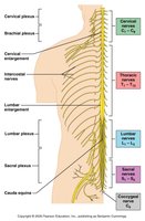
There are 31 pairs of spinal nerves:
8 pairs of cervical nerves (C1–C8)
12 pairs of thoracic nerves (T1–T12)
5 pairs of lumbar nerves (L1–L5)
5 pairs of sacral nerves (S1–S5)
1 pair of coccygeal nerves (Co1)
Cervical nerves emerge above their corresponding vertebrae (except C8, which emerges between C7 and T1); all others emerge below their vertebrae.
Nerve Plexuses
A nerve plexus is a complex, interwoven network of nerves formed by the ventral rami of spinal nerves. There are four major plexuses:
Cervical Plexus (C1–C5): Innervates the neck and diaphragm (phrenic nerve).
Brachial Plexus (C5–T1): Innervates the shoulder, arm, and hand (major nerves: median, ulnar, radial).
Lumbar Plexus (L1–L4): Innervates the lower abdomen and anterior thigh (major nerve: femoral).
Sacral Plexus (L4–S4): Innervates the posterior thigh, leg, and foot (major nerve: sciatic).
Major Peripheral Nerves
Phrenic Nerve: Arises from the cervical plexus (C3–C5); innervates the diaphragm.
Radial, Median, Ulnar Nerves: Arise from the brachial plexus; supply muscles and skin of the upper limb.
Femoral Nerve: Arises from the lumbar plexus; supplies anterior thigh muscles.
Sciatic Nerve: Arises from the sacral plexus; the largest nerve in the body, supplies the posterior thigh, leg, and foot.
Clinical Relevance
Lumbar Puncture (Spinal Tap)
Performed below L3 to avoid injury to the spinal cord (which ends at L1–L2 in adults).
Used to collect CSF for diagnostic purposes (e.g., infection detection).
Summary Table: Spinal Cord Anatomy in Cross Section
Region | Structure | Function |
|---|---|---|
White Matter | Columns (funiculi) | Conducts sensory and motor signals |
Gray Matter | Horns (anterior, posterior, lateral) | Integration of sensory and motor information |
Central Canal | CSF-filled cavity | Cushions and nourishes spinal cord |
Dorsal Root | Sensory fibers | Transmits sensory input to spinal cord |
Ventral Root | Motor fibers | Transmits motor output from spinal cord |
Key Terms and Definitions
Conus Medullaris: Tapered end of the spinal cord.
Cauda Equina: Bundle of nerve roots below the conus medullaris.
Filum Terminale: Fibrous extension of pia mater anchoring the spinal cord to the coccyx.
Meninges: Three protective membranes (dura mater, arachnoid mater, pia mater) surrounding the spinal cord.
Plexus: Network of intersecting nerves.
Additional info:
The spinal cord is organized segmentally, with each segment giving rise to a pair of spinal nerves.
Damage to specific regions of the spinal cord or its nerves can result in characteristic patterns of sensory or motor loss, aiding in clinical diagnosis.
