 Back
BackStudy Guide: The Respiratory System (Anatomy & Physiology)
Study Guide - Smart Notes
 Tailored notes based on your materials, expanded with key definitions, examples, and context.
The Respiratory System: Structural Plan and Overview
Basic Structural Plan
The respiratory system is organized similarly to an inverted tree, with the alveoli functioning as the 'leaves' where gas exchange occurs. The system is composed of a series of branching tubes that culminate in microscopic air sacs. - Alveoli: Microscopic sacs surrounded by capillaries, essential for gas exchange via diffusion. - Diffusion: The primary mechanism for gas exchange in the respiratory system. 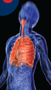
Major Divisions of the Respiratory Tract
The respiratory tract is divided into upper and lower regions, each with distinct anatomical structures and functions. - Upper respiratory tract: Nose, pharynx, larynx - Lower respiratory tract: Trachea, bronchial tree, lungs 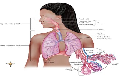
Respiratory Mucosa
Structure and Function
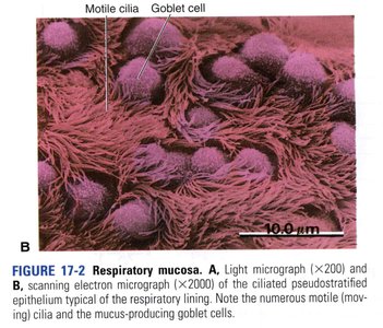
The respiratory mucosa is a specialized mucous membrane lining the airways, playing a crucial role in air purification and protection. - Mucus blanket: Over 125 mL of mucus produced daily traps dust and pollen. - Cilia: Beat in one direction to move mucus upward for removal. - Goblet cells: Produce mucus; found in pseudostratified epithelium. 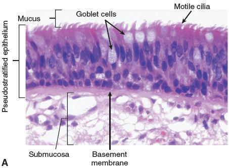
Anatomy of the Nose and Paranasal Sinuses
Nose Structure
The nose is divided by the nasal septum and lined with mucous membrane. It contains sinuses that drain into the nasal cavity. - Nasal polyps: Noncancerous growths associated with chronic hay fever. - Sinuses: Frontal, maxillary, sphenoidal, ethmoidal.
Functions of the Nose
- Air conditioning: Warms and moistens inhaled air. - Olfaction: Contains sense organs for smell.
Paranasal Sinuses
- Sinuses: Air-filled spaces that lighten the skull and aid in voice resonance. 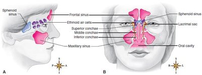
Pharynx (Throat)
Structure
The pharynx is a muscular tube about 12.5 cm long, divided into three regions: nasopharynx, oropharynx, and laryngopharynx. It serves as a passageway for both air and food. 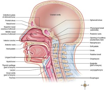
Functions
- Passageway: For food, liquids, and air. - Immune protection: Tonsils embedded in the pharynx provide defense against pathogens.
Tonsillitis
- Tonsillitis: Inflammation of the tonsils, often visible as swollen palatine tonsils. 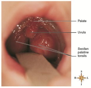
Larynx (Voice Box)
Structure
The larynx is located below the pharynx and is composed of several cartilages, including the thyroid cartilage (Adam's apple) and the epiglottis. - Vocal cords: Stretch across the interior, forming the glottis. 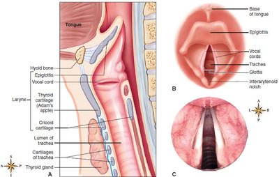
Functions
- Air passage: Directs air to and from the lungs. - Voice production: Vibrations of the vocal cords produce sound.
Trachea (Windpipe)
Structure
The trachea is a tube about 11 cm long, supported by C-shaped rings of cartilage to prevent collapse. It extends from the larynx into the thoracic cavity. 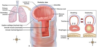
Function
- Air passage: Conducts air to and from the lungs.
Clinical Applications
- Obstruction: Blockage can be fatal; abdominal thrusts (Heimlich maneuver) and tracheostomy are emergency interventions. 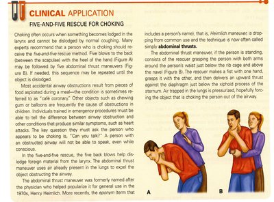
Bronchi, Bronchioles, and Alveoli
Structure
The trachea branches into right and left primary bronchi, which further divide into secondary bronchi and bronchioles, ending in clusters of alveolar sacs. 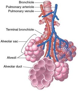
Functions
- Bronchi and bronchioles: Distribute air to alveoli. - Alveoli: Site of gas exchange; type II cells produce surfactant to reduce surface tension. 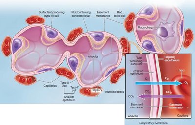
Lungs and Pleura
Structure of the Lungs
The lungs are large organs filling the chest cavity, except for the space occupied by the heart. Each lung has an apex (top) and base (bottom). 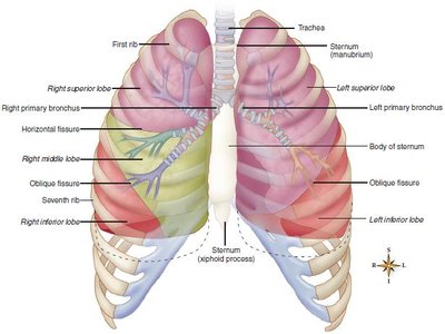
Pleura
The pleura is a moist, slippery membrane covering the lungs and lining the chest cavity, reducing friction during breathing.
Pleurisy and Atelectasis
- Pleurisy: Inflammation of the pleura. - Atelectasis: Collapse of lung tissue, often due to pneumothorax (air in pleural space) or hemothorax (blood in pleural space). 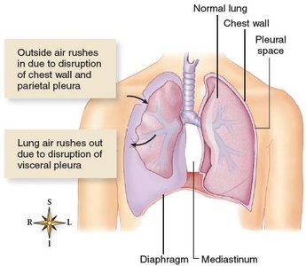
Conditions of the Lower Respiratory Tract
Common Disorders
- Acute bronchitis: Inflammation of the bronchi. - Pneumonia: Infection causing alveoli and bronchi to fill with exudate. - Tuberculosis: Chronic infection caused by Mycobacterium tuberculosis. - Restrictive pulmonary disorders: Limit lung expansion. - Obstructive pulmonary disorders: Include COPD, chronic bronchitis, emphysema, asthma. - Lung cancer: Malignant growth in lung tissue. 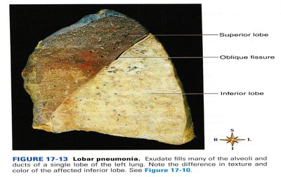
Mechanics of Breathing (Pulmonary Ventilation)
Phases of Breathing
Breathing consists of inspiration (air in) and expiration (air out), driven by changes in thoracic volume and pressure. - Inspiration: Active process; diaphragm and external intercostals contract, increasing thoracic volume and decreasing pressure. - Expiration: Passive process; thorax returns to resting size, increasing pressure and expelling air. Forceful expiration uses internal intercostals and abdominal muscles. 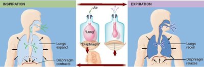
Volumes of Air Exchanged
Pulmonary ventilation volumes are measured with a spirometer.
Volume | Description |
|---|---|
Tidal Volume (TV) | Amount normally breathed in or out with each breath |
Vital Capacity (VC) | Largest amount of air exhaled in one expiration |
Expiratory Reserve Volume (ERV) | Amount forcibly exhaled after tidal volume |
Inspiratory Reserve Volume (IRV) | Amount forcibly inhaled after normal inspiration |
Residual Volume (RV) | Air remaining after forceful expiration |
Regulation of Ventilation
Neural Control
Breathing is regulated by respiratory control centers in the brainstem (medulla and pons). - Medullary rhythmicity area: Sets basic rate and depth (12-18 breaths/min). - Pontine centers: Adjust breathing rhythm as needed.
Influences on Respiration
- Cerebral cortex: Allows voluntary (limited) control. - Chemoreceptors: Respond to CO2, O2, and pH changes. - Pulmonary stretch receptors: Prevent overinflation. 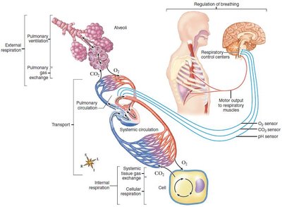
Breathing Patterns
Types of Breathing
- Eupnea: Normal breathing. - Hyperventilation: Rapid, deep breathing. - Hypoventilation: Slow, shallow breathing. - Dyspnea: Difficult or labored breathing. - Orthopnea: Dyspnea relieved by upright position. - Apnea: Cessation of breathing. - Cheyne-Stokes respiration: Alternating apnea and hyperventilation. - Respiratory arrest: Failure to resume breathing after apnea.
Pulmonary Gas Exchange and Transport
Gas Exchange in the Lungs
- Oxygen: Moves from alveoli to capillaries, binds hemoglobin to form oxyhemoglobin. - Carbon dioxide: Moves from capillaries to alveoli, expelled in expired air.
Transport of Gases in Blood
Gas | Transport Form | Percentage |
|---|---|---|
Oxygen (O2) | Oxyhemoglobin (HbO2) | Most |
Carbon Dioxide (CO2) | Dissolved | 10% |
Carbon Dioxide (CO2) | Carbaminohemoglobin (HbCO2) | 20% |
Carbon Dioxide (CO2) | Bicarbonate ions (HCO3-) | 70% |
Key Equations
Gas Laws and Pressure Gradients
- Boyle's Law: (Pressure and volume are inversely related) - Pressure Gradient: Air moves from high to low pressure.
Summary Table: Respiratory Structures and Functions
Structure | Main Function |
|---|---|
Nose | Warms, moistens air; olfaction |
Pharynx | Passageway for air/food; immune protection |
Larynx | Air passage; voice production |
Trachea | Air passage; prevents collapse |
Bronchi/Bronchioles | Air distribution |
Alveoli | Gas exchange |
Pleura | Reduces friction |
Clinical Applications and Review Questions
Clinical Applications
- Endotracheal intubation: Tube placed to maintain airway. - Tracheostomy: Surgical opening for breathing. - Abdominal thrust maneuver: Emergency technique for airway obstruction.
Review Questions
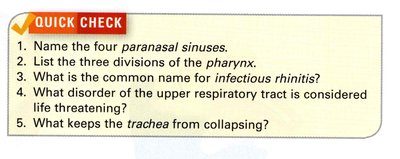
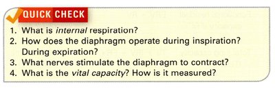
- What are the primary functions of the respiratory system? - Describe the characteristics of the alveoli that enable them to perform their function of gas exchange. - What distinguishes the upper respiratory tract from the lower respiratory tract? - What is the role of the respiratory mucosa? - Name the four paranasal sinuses. - List the three divisions of the pharynx. - What keeps the trachea from collapsing? - What lung structures serve to distribute air, and which serve as gas exchangers? - Describe the function of surfactant. - What causes pleurisy? - How does a pneumothorax differ from a hemothorax? - What is the difference between bronchopneumonia and lobar pneumonia? - Give two examples of chronic obstructive pulmonary disease (COPD). - What is internal respiration? - How does the diaphragm operate during inspiration and expiration? - What nerves stimulate the diaphragm to contract? - What is the vital capacity? How is it measured? Additional info: Academic context was added to clarify mechanisms, clinical relevance, and to provide self-contained explanations for each topic.
