 Back
BackStudy Guide: The Special Senses in Anatomy & Physiology
Study Guide - Smart Notes
 Tailored notes based on your materials, expanded with key definitions, examples, and context.
The Special Senses
Overview of the Special Senses
The special senses include olfaction (smell), gustation (taste), vision, hearing, and equilibrium. These senses are mediated by specialized sensory organs and pathways that allow the human body to detect and interpret environmental stimuli.
Olfaction: Detection of airborne chemicals by the olfactory epithelium in the nasal cavity.
Gustation: Detection of dissolved chemicals by taste buds on the tongue.
Vision: Detection of light by photoreceptors in the retina of the eye.
Hearing: Detection of sound waves by the cochlea in the inner ear.
Equilibrium: Detection of head position and movement by the vestibular apparatus in the inner ear.
Olfaction (Smell)
Anatomy of Olfactory Organs
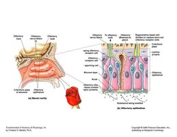
Olfactory organs are located in the nasal cavity, on either side of the nasal septum. The olfactory epithelium contains specialized cells responsible for detecting odorants.
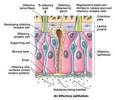
Olfactory epithelium: Contains olfactory receptor cells, supporting cells, and basal cells.
Olfactory receptor cells: Neurons with cilia that bind odor molecules.
Supporting cells: Provide structural and metabolic support.
Basal cells: Regenerate olfactory receptor cells.
Olfactory Pathways to the Brain
The olfactory pathway transmits sensory information from the nasal cavity to the brain.
Axons of olfactory receptor cells collect into bundles and penetrate the cribriform plate of the ethmoid bone.
These axons reach the olfactory bulbs, where the first synapse occurs.
Axons leaving the olfactory bulb travel along the olfactory tract to reach the olfactory cortex, hypothalamus, and portions of the limbic system.
Olfactory information reaches processing centers without first synapsing in the thalamus.
Gustation (Taste)
Anatomy of Taste Buds and Lingual Papillae
Taste buds are sensory organs associated with epithelial projections called lingual papillae on the dorsal surface of the tongue.
Filiform papillae: Provide friction, do not contain taste buds.
Fungiform papillae: Contain about 5 taste buds each.
Circumvallate papillae: Contain about 100 taste buds each.
Elements of Taste Perception
Sweet
Salty
Bitter
Sour
Umami
Gustatory Pathways to the Brain
Taste buds are monitored by cranial nerves that synapse within the solitary nucleus of the medulla oblongata, then relay information to the thalamus and primary sensory cortex.
Cranial nerves involved: Facial nerve (VII) and glossopharyngeal nerve (IX).
Taste Sensitivity
Exhibits significant individual differences.
Some conditions are inherited, such as the ability to taste phenylthiocarbamide (PTC).
Vision
Accessory Structures of the Eye
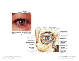
The accessory structures of the eye protect and maintain the function of the eyeball.
Eyelids (palpebrae): Continuation of skin; blinking lubricates and cleans the eye.
Medial and lateral canthus: Points where eyelids connect.
Lacrimal caruncle: Contains glands producing thick secretions.
Conjunctiva: Epithelium covering inner surfaces of eyelids and outer surface of eye.
Lacrimal apparatus: Produces, distributes, and removes tears.
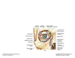
Internal Structures of the Eye
The eyeball is hollow and divided into two cavities: a large posterior cavity and a smaller anterior cavity.
Orbital fat: Cushions and insulates the eye.
Fibrous tunic: The most superficial layer, includes sclera, cornea, and limbus.
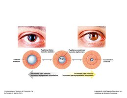
Vascular tunic (uvea): Middle layer, includes iris, ciliary body, and choroid.
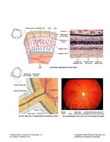
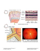
Neural tunic (retina): Deepest layer, contains photoreceptors (rods and cones).
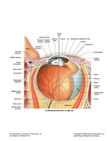
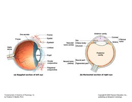
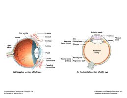
Functions of Eye Layers
Fibrous tunic: Provides protection and shape.
Vascular tunic: Supplies blood, regulates light, secretes aqueous humor, controls lens shape.
Neural tunic: Contains rods (sensitive to light, black-and-white vision) and cones (color vision, high resolution).
Chambers and Fluids of the Eye
Anterior cavity: Subdivided into anterior chamber (cornea to iris) and posterior chamber (iris, ciliary body, lens).
Aqueous humor: Fluid circulating within the eye, maintains intraocular pressure.
Canal of Schlemm: Drains aqueous humor back into circulation.
Pathologies
Cataract: Loss of lens transparency.
Glaucoma: Abnormally high intraocular pressure.
Light Refraction and Accommodation
Light refraction: Bending of light by cornea and lens.
Accommodation: Shape of lens changes to focus image on retina.
Visual Pathway
The visual pathway begins at photoreceptors and ends at the visual cortex of the cerebral hemispheres.
Photoreceptors (rods and cones) detect light.
Signals are transmitted via the optic nerve to the brain.
Hearing and Equilibrium
Anatomy of the Ear
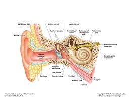
The ear is divided into three regions: external ear, middle ear, and inner ear.
External ear: Auricle (pinna), external acoustic canal, tympanic membrane.
Middle ear: Tympanic cavity, auditory tube, three auditory ossicles (malleus, incus, stapes).
Inner ear: Bony labyrinth, membranous labyrinth, vestibule, semicircular canals, cochlea.
Functions of Ear Structures
Auricle: Directs sound waves into the external acoustic canal.
Tympanic membrane: Converts sound waves into mechanical movements.
Ceruminous glands: Produce cerumen (earwax) to protect the ear.
Auditory ossicles: Conduct vibrations to the inner ear.
Tensor tympani and stapedius muscles: Protect the ear from loud sounds by stiffening the tympanic membrane and reducing stapes movement.
Inner Ear and Equilibrium
Vestibule: Contains saccule and utricle; detects gravity and linear acceleration.
Semicircular canals: Detect rotational movements of the head.
Cochlea: Detects sound waves.
Hearing Process
Hearing involves the conversion of sound waves into electrical signals interpreted by the brain.
Sound waves enter the external acoustic canal.
Tympanic membrane vibrates.
Auditory ossicles transmit vibrations to the oval window.
Pressure waves are created in the perilymph of the cochlea.
Hair cells in the cochlear duct are stimulated.
Signals are sent to the auditory cortex via the cochlear nerve.
Sound Properties
Frequency: Determined by which part of the cochlear duct is stimulated.
Intensity (volume): Determined by the number of hair cells stimulated.
Sound energy: Measured in decibels (dB).
Aging Effects on Hearing
Tympanic membrane becomes less flexible.
Articulations between ossicles stiffen.
Round window may ossify, reducing hearing ability.
Summary Table: Major Structures and Functions of the Special Senses
Sense | Main Organ | Key Structures | Function |
|---|---|---|---|
Olfaction | Nasal cavity | Olfactory epithelium, olfactory bulbs | Detects airborne chemicals |
Gustation | Tongue | Taste buds, lingual papillae | Detects dissolved chemicals |
Vision | Eye | Cornea, lens, retina | Detects light |
Hearing | Ear | Cochlea, auditory ossicles | Detects sound waves |
Equilibrium | Ear | Vestibule, semicircular canals | Detects head position and movement |
Additional info: Academic context was added to clarify the pathways, functions, and clinical relevance of each special sense. The notes are structured to provide a comprehensive yet concise review for exam preparation.
