 Back
BackStudy Notes: Functional Anatomy of the Respiratory System
Study Guide - Smart Notes
 Tailored notes based on your materials, expanded with key definitions, examples, and context.
Functional Anatomy of the Respiratory System
Overview of the Respiratory System
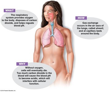
The respiratory system is responsible for gas exchange between the blood and the external environment. Its major structures include the nose, pharynx, larynx, trachea, bronchi, and lungs (alveoli). The system provides oxygen to the body, disposes of carbon dioxide, and helps regulate blood pH.
Gas exchange occurs in the air sacs of the lungs, called alveoli, and at capillary beds around the body.
Oxygen is vital for cellular respiration and ATP production.
Carbon dioxide is a waste product of cellular respiration and must be removed to prevent blood acidity.
Protective Mechanisms of the Respiratory System
The respiratory system employs several protective (immune) mechanisms to ensure that the air reaching the lungs is clean, warm, and moist.
Mucous membrane (mucosa): Lines the respiratory passages, secretes surfactant, and traps irritants.
Hairs and mucus: Remove dust and bacteria from incoming air.
Ciliated cells: Move contaminated mucus toward the throat for swallowing and digestion.
Lysozyme enzymes: Destroy bacteria chemically in the mucus.
Alveolar macrophages: Patrol and protect alveoli from infection; also called "dust cells".
Respiratory Passageway: From Nasal Cavity to Alveoli
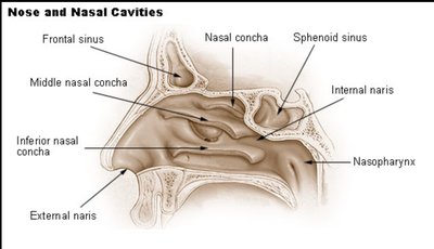
Nose and Nasal Cavities
The nose is the only external organ of the respiratory system. Air enters through the nostrils (nares) into the nasal cavity, which is lined with hair and mucus. The nasal cavity is divided by the nasal septum and contains mucosa-covered projections called conchae that increase surface area and turbulence, trapping particles.
Paranasal sinuses: Surround the nasal cavity, act as resonance chambers for speech, and lighten the skull.
Palate: Separates the nose from the mouth; cleft palate is a genetic defect affecting breathing and speech.
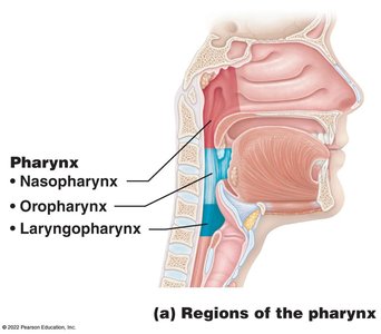
Pharynx (Throat)
The pharynx is a muscular passageway shared by the respiratory and digestive systems. It connects the nasal cavity to the larynx and esophagus and is divided into three regions:
Nasopharynx: Behind the nose; carries air only, lined with mucus and blood vessels.
Oropharynx: Behind the mouth; common passage for food and air.
Laryngopharynx: Throat region; common passage for food and air.
Tonsils are clusters of lymphatic tissue surrounding the pharynx, providing immune protection.
Larynx (Voice Box)
The larynx routes air and plays a role in speech. It contains vocal cords, which vibrate to produce sound. The larynx is formed by rigid cartilages, including the thyroid cartilage (Adam’s apple) and the epiglottis, which covers the tracheal opening during swallowing to prevent food from entering the airway.
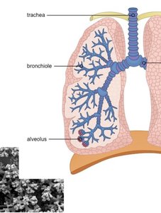
Trachea (Windpipe)
The trachea connects the larynx to the bronchi. It is lined with ciliated mucosa and contains rings of hyaline cartilage to keep it open. Goblet cells produce mucus, and cilia push mucus toward the throat for digestion.
Structure and Function of the Lungs and Pleural Coverings
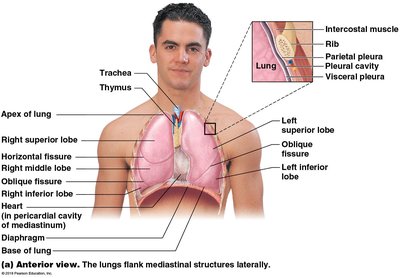
Lungs
The lungs are organs of breathing located on either side of the mediastinum. They are composed mainly of elastic connective tissue and are divided into lobes:
Right lung: Shorter, heavier, three lobes (superior, middle, inferior).
Left lung: Longer, lighter, two lobes (superior, inferior) to accommodate the heart.
Each lung is covered by a serous membrane called the pleura:
Visceral pleura: Attached to the lung surface.
Parietal pleura: Lines the thoracic cavity.
Pleural fluid: Allows membranes to slide past each other with minimal friction.
Bronchi and Bronchioles
The bronchi are formed by the division of the trachea and connect to each lung. Inside the lungs, bronchi divide into smaller airways called bronchioles, which greatly increase the surface area for gas exchange. Each bronchiole terminates in a cluster of air sacs called alveoli.
Respiratory Zone: Gas Exchange Structures
Respiratory Zone Structures
The respiratory zone is where gas exchange occurs. Its structures include:
Respiratory bronchioles
Alveolar ducts
Alveolar sacs
Alveoli
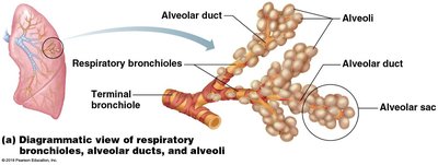
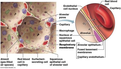
Alveoli and Gas Exchange
Alveoli are small pouches clustered together to form alveolar sacs. Each alveolus has a cavity and a wall composed of a single layer of epithelial cells. The wall expands and contracts with inhalation and exhalation. Alveoli provide a large surface area (~70 m2) for gas exchange.
Cell types in alveoli:
Simple squamous epithelial cells (wall structure)
Simple cuboidal surfactant-secreting cells
Alveolar macrophages (immune defense)
Alveolar pores (connect neighboring air sacs)
Surfactant: Lipoprotein secretion that decreases surface tension, facilitates inflation/deflation, and provides immune function.
Pulmonary capillaries: Surround alveoli, walls are one cell thick for rapid gas exchange.
Stroma: Elastic connective tissue allowing lung recoil.
Respiratory Membrane (Air-Blood Barrier)
The respiratory membrane is formed by the alveolar and capillary walls. On one side is air, and on the other is blood. Gas crosses the membrane by diffusion:
Oxygen: Enters the blood from the alveoli.
Carbon dioxide: Enters the alveoli from the blood.
Key blood vessels:
Pulmonary arteriole: Carries deoxygenated blood from the heart to the alveoli.
Pulmonary venule: Carries oxygenated blood from the alveoli to the heart.
Order of Respiratory Passageway
The order of structures from the site where air enters the nostrils to the end passages of the lungs:
Nasal cavity
Pharynx
Larynx
Trachea
Bronchi
Bronchioles
Alveoli
Summary Table: Respiratory Zone Structures
Structure | Main Function |
|---|---|
Respiratory bronchiole | Conducts air, begins gas exchange |
Alveolar duct | Passageway to alveolar sacs |
Alveolar sac | Cluster of alveoli, site of gas exchange |
Alveolus | Individual air sac, gas exchange with capillaries |
Key Terms and Definitions
Alveoli: Air sacs where gas exchange occurs.
Bronchi: Main passageways into the lungs.
Bronchioles: Smaller branches of the bronchi.
Pharynx: Shared passageway for air and food.
Larynx: Voice box, routes air and produces sound.
Trachea: Windpipe, connects larynx to bronchi.
Pleura: Serous membrane covering the lungs.
Surfactant: Substance reducing surface tension in alveoli.
Respiratory membrane: Barrier for gas exchange between air and blood.
Relevant Equations
Gas exchange across the respiratory membrane occurs by diffusion, governed by partial pressure gradients:
Where is the difference in partial pressure, is the partial pressure in the alveoli, and is the partial pressure in the capillaries.
Oxygen and carbon dioxide move from areas of higher concentration to lower concentration:
Additional info:
Some details about the immune function of surfactant and the role of elastic tissue in lung recoil were inferred for completeness.
