 Back
BackSystemic Blood Flow and Major Vascular Pathways
Study Guide - Smart Notes
 Tailored notes based on your materials, expanded with key definitions, examples, and context.
Systemic Blood Flow: Overview
General Systemic Flow from the Heart
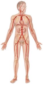
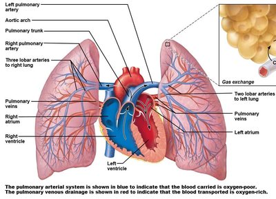
The systemic circulation is responsible for delivering oxygenated blood from the left ventricle of the heart to all body tissues and returning deoxygenated blood to the right atrium. The aorta is the main artery that branches to supply all regions of the body.
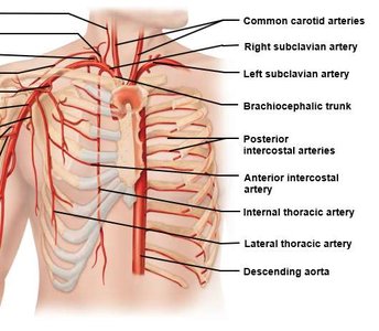
Aortic Arch Branches: The three major branches are the brachiocephalic trunk, left common carotid artery, and left subclavian artery.
Descending Aorta: Continues as the thoracic and abdominal aorta, giving rise to arteries that supply the thorax, abdomen, pelvis, and limbs.
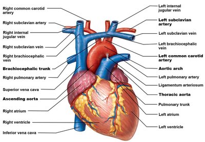
Venous Drainage: Superior vs. Inferior Vena Cava
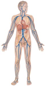
Venous blood from the body returns to the heart via two large veins:
Superior Vena Cava (SVC): Drains blood from the head, neck, upper limbs, and thoracic wall.
Inferior Vena Cava (IVC): Drains blood from the abdomen, pelvis, and lower limbs.
Blood Flow to and from the Head & Neck
Arterial Supply
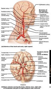
Blood is supplied to the head and neck by paired arteries branching from the aortic arch and subclavian arteries.
Common Carotid Arteries: Each splits into external (supplies face and scalp) and internal (supplies brain) carotid arteries.
Subclavian Arteries: Give rise to vertebral arteries (supply brain), thyrocervical trunk, and costocervical trunk.
Cerebral Arterial Circle (Circle of Willis): Formed by anastomoses of vertebral and internal carotid arteries, ensuring continuous brain perfusion.
Venous Drainage
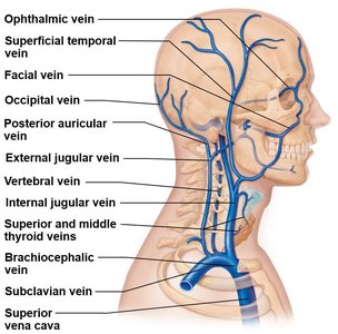
Venous blood from the head and neck drains through three major paired routes:
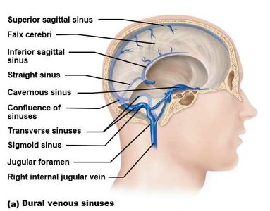
Internal Jugular Veins: Drain the brain via dural venous sinuses, exit skull through jugular foramen.
External Jugular Veins: Drain superficial structures, empty into subclavian vein.
Vertebral Veins: Drain cervical spinal cord and posterior skull, descend through transverse foramina.
Blood Flow to and from the Thoracic Wall and Organs
Thoracic Wall Arterial Supply
The thoracic wall receives blood from branches of the thoracic aorta and subclavian arteries.
Posterior Intercostal Arteries: Supply posterior thoracic wall.
Internal Thoracic Artery: Branches from subclavian, supplies anterior thoracic wall via anterior intercostal arteries.
Thoracic Organ Supply and Drainage
Bronchial Arteries: Supply oxygenated blood to lung tissue.
Other Branches: Small arteries supply esophagus, mediastinum, and pericardium.
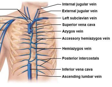
Venous Drainage: Blood from thoracic organs drains into the azygos system, which empties into the SVC.
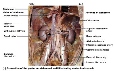
Blood Flow to and from the Abdominal Wall and Organs
Branches of the Abdominal Aorta
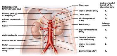
The abdominal aorta gives rise to paired and unpaired branches supplying the abdominal wall and organs.
Paired Branches: Inferior phrenic, middle suprarenal, renal, and gonadal arteries supply diaphragm, adrenal glands, kidneys, and gonads, respectively.
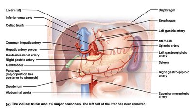
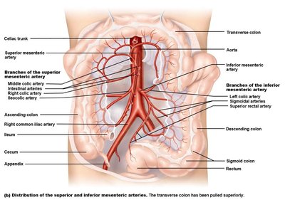
Unpaired (Midline) Branches: Celiac trunk, superior mesenteric artery, and inferior mesenteric artery supply the gastrointestinal tract and accessory organs.
Blood Flow to and from the Pelvis
Pelvic Arterial Supply
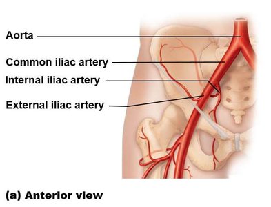
At the level of L4, the abdominal aorta bifurcates into the common iliac arteries, which further divide into internal and external iliac arteries.
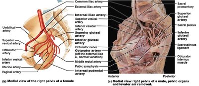
Internal Iliac Artery: Supplies pelvic organs, gluteal muscles, perineum, and adductor muscles via branches such as the superior/inferior gluteal, internal pudendal, and obturator arteries.
External Iliac Artery: Continues as the femoral artery to supply the lower limb.
Hepatic Portal System
Structure and Function
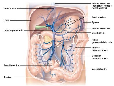
The hepatic portal system is a specialized venous network that directs blood from the gastrointestinal tract and spleen to the liver for processing before it enters the systemic circulation.
Hepatic Portal Vein: Formed by the union of the splenic, superior mesenteric, and inferior mesenteric veins; carries nutrient-rich blood to the liver.
Hepatic Veins: Drain processed blood from the liver into the inferior vena cava.
Venous Drainage from Abdominal and Pelvic Organs
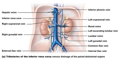
Venous blood from abdominal and pelvic organs returns to the heart via the inferior vena cava, which receives blood from lumbar, renal, hepatic, gonadal, and adrenal veins.
Right Gonadal and Adrenal Veins: Drain directly into the IVC.
Left Gonadal and Adrenal Veins: Drain into the left renal vein before reaching the IVC.
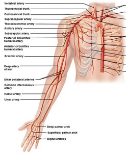
Blood Flow to and from the Upper Limb
Arterial Supply
The upper limb is supplied by branches of the subclavian artery, which becomes the axillary, brachial, radial, and ulnar arteries as it passes through the limb.
Brachial Artery: Main artery of the arm, commonly used for blood pressure measurement.
Radial and Ulnar Arteries: Supply the forearm and hand, forming palmar arches.
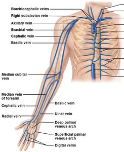
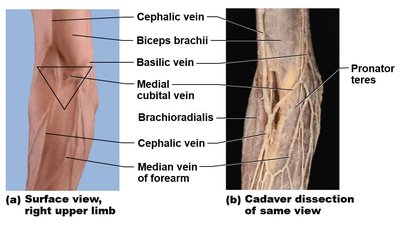
Venous Drainage
Deep Veins: Accompany arteries and drain into the axillary and subclavian veins.
Superficial Veins: Visible through the skin, commonly used for venipuncture (e.g., median cubital vein).
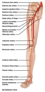
Blood Flow to and from the Lower Limb
Arterial Supply
The external iliac artery becomes the femoral artery as it enters the thigh, supplying the lower limb through its branches.
Femoral Artery: Main artery of the thigh, palpable at the inguinal ligament.
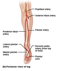
Popliteal Artery: Continuation of the femoral artery behind the knee, branching into anterior and posterior tibial arteries.
Tibial and Fibular Arteries: Supply the leg and foot, forming the plantar arch.
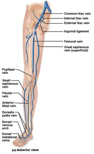
Venous Drainage
Deep Veins: Follow the arteries, draining into the femoral and external iliac veins.
Superficial Veins: Include the great and small saphenous veins, important for venous return and clinical procedures.
Fetal versus Postnatal Circulation
Specialized Fetal Structures
Fetal circulation includes unique structures that bypass nonfunctional fetal lungs and direct oxygenated blood from the placenta to the systemic circulation.
Placenta: Site of gas and nutrient exchange between maternal and fetal blood.
Umbilical Vessels: Umbilical vein carries oxygenated blood to fetus; umbilical arteries return deoxygenated blood to placenta.
Ductus Venosus: Shunts blood from umbilical vein to inferior vena cava, bypassing the liver.
Foramen Ovale: Opening between right and left atria, allowing blood to bypass fetal lungs.
Ductus Arteriosus: Connects pulmonary trunk to aorta, further bypassing the lungs.
At birth, these shunts close, and normal adult circulation is established.
