 Back
BackThe Auditory System and Balance: Structure and Function of the Ear
Study Guide - Smart Notes
 Tailored notes based on your materials, expanded with key definitions, examples, and context.
The Ear: Hearing and Balance
Overview of Auditory and Equilibrium Functions
The ear is a complex organ responsible for both hearing and equilibrium (balance). Although the organs for these senses are structurally connected, their receptors respond to different stimuli and are activated independently. The movement of fluids within the ear is essential for stimulating mechanoreceptors that enable these senses.
Hearing: Detection of sound waves and their conversion into neural signals.
Equilibrium: Maintenance of balance and spatial orientation.
What is Sound?
Physical Properties of Sound
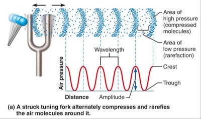
Sound is produced by vibrating objects, which create alternating areas of high and low pressure in the surrounding air. These pressure changes propagate as waves, which are detected by the ear.
Wavelength: The distance between two consecutive crests or troughs of a sound wave.
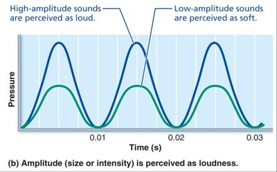
Amplitude: The height of the wave, which determines the loudness of the sound.
Frequency: The number of waves passing a point per second, perceived as pitch.
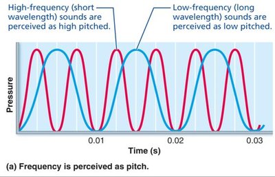
High-frequency (short wavelength) sounds are perceived as high-pitched.
Low-frequency (long wavelength) sounds are perceived as low-pitched.
High-amplitude sounds are perceived as loud, while low-amplitude sounds are soft.
Anatomy of the Ear
Major Regions of the Ear
The ear is divided into three major regions, each with specialized structures and functions:
External (Outer) Ear: Involved in hearing only.
Middle Ear (Tympanic Cavity): Involved in hearing only.
Internal (Inner) Ear: Involved in both hearing and equilibrium.
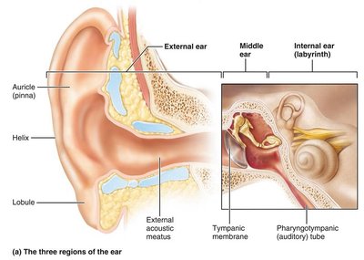
External Ear
Auricle (Pinna): Shell-shaped structure that funnels sound waves into the auditory canal. Includes the helix (cartilaginous rim) and lobule (fleshy earlobe).
External Acoustic Meatus (Auditory Canal): Short, curved tube lined with skin, hairs, sebaceous glands, and ceruminous (earwax) glands. Transmits sound waves to the tympanic membrane.
Tympanic Membrane (Eardrum): Thin, translucent connective tissue membrane that vibrates in response to sound and transfers sound energy to the middle ear bones.
Middle Ear
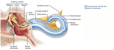
The middle ear is an air-filled cavity within the temporal bone, bordered laterally by the eardrum and medially by the oval and round windows.
Pharyngotympanic (Auditory) Tube: Connects the middle ear to the nasopharynx, allowing pressure equalization across the tympanic membrane.
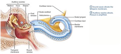
Auditory Ossicles: Three small bones (malleus, incus, stapes) that transmit and amplify vibrations from the tympanic membrane to the oval window.
Muscles: Tensor tympani and stapedius muscles contract reflexively to protect hearing receptors from loud sounds.
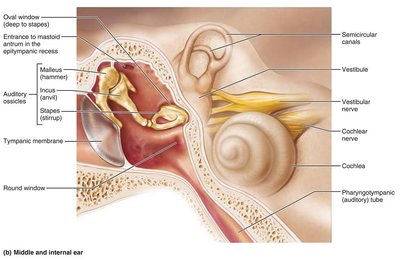

Internal Ear (Labyrinth)
The internal ear consists of a bony labyrinth filled with perilymph and a membranous labyrinth filled with endolymph. It is divided into three regions: vestibule, semicircular canals, and cochlea.
Bony Labyrinth: System of channels in the bone, filled with perilymph.
Membranous Labyrinth: Series of sacs and ducts within the bony labyrinth, filled with potassium-rich endolymph.
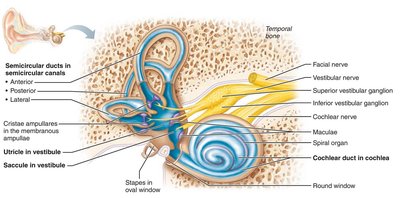
Vestibule
Central egg-shaped cavity containing the saccule and utricle, which house equilibrium receptors (maculae) that respond to gravity and changes in head position.
Semicircular Canals
Three canals oriented in different planes, each containing a membranous semicircular duct and an enlarged region called the ampulla, which houses the crista ampullaris (receptor for rotational movements).
Cochlea
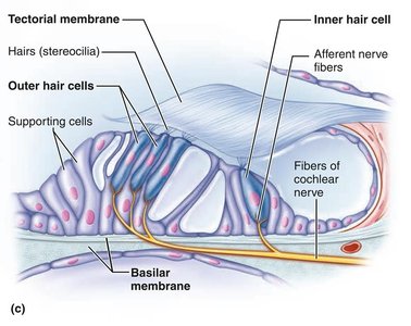
Spiral, conical chamber that contains the cochlear duct, which houses the spiral organ (organ of Corti)—the receptor organ for hearing.
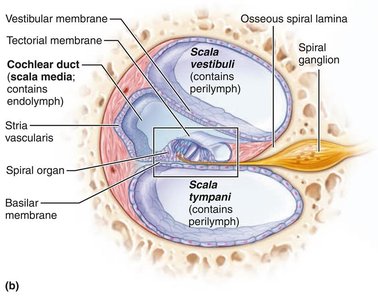
Chambers of the Cochlea
Scala Vestibuli: Contains perilymph and abuts the oval window.
Scala Media (Cochlear Duct): Contains endolymph and houses the spiral organ.
Scala Tympani: Contains perilymph and terminates at the round window.

Summary Table: Internal Ear Structures and Functions
Bony Labyrinth | Membranous Labyrinth | Function | Receptor Region |
|---|---|---|---|
Semicircular canals | Semicircular ducts | Equilibrium: rotational (angular) acceleration | Crista ampullaris |
Vestibule | Utricle and saccule | Equilibrium: head position relative to gravity, linear acceleration | Macula |
Cochlea | Cochlear duct (scala media) | Hearing | Spiral organ |
Pathway of Sound Waves Through the Ear
Transmission of Sound
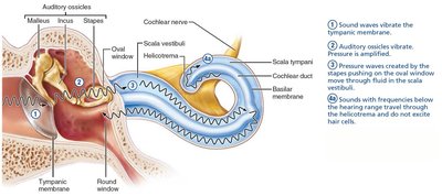
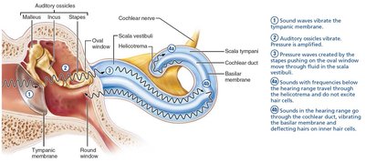
Sound waves enter the external acoustic meatus and vibrate the tympanic membrane.
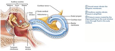
Vibrations are transferred to the auditory ossicles, which amplify the pressure and transmit it to the oval window.
Stapes movement at the oval window creates pressure waves in the perilymph of the scala vestibuli.
Waves with frequencies below the threshold of hearing travel through the helicotrema and do not excite hair cells.
Sounds within the hearing range travel through the cochlear duct, vibrating the basilar membrane and deflecting hairs on inner hair cells.
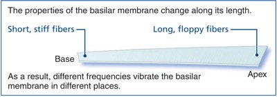
Resonance of the Basilar Membrane
The basilar membrane varies in structure along its length, allowing it to resonate at different frequencies:
Base (near oval window): Short, stiff fibers resonate with high-frequency waves.
Apex (cochlear tip): Long, floppy fibers resonate with low-frequency waves.

Sound Transduction
Excitation of Hair Cells
Movement of the basilar membrane deflects the stereocilia of inner hair cells, which are embedded in the tectorial membrane. This mechanical deflection opens or closes ion channels, leading to changes in the hair cell's membrane potential and neurotransmitter release.
Bending toward tallest stereocilia: Opens K+ and Ca2+ channels, causing depolarization.
Bending toward shortest stereocilia: Closes channels, causing hyperpolarization.
Role of Outer Hair Cells
Outer hair cells can contract and stretch, changing the stiffness of the basilar membrane.
This amplifies the responsiveness of inner hair cells and protects them from loud noises by decreasing basilar membrane motion.
Auditory Pathway and Processing
Neural Pathway
Auditory information is transmitted from the cochlear receptors (inner hair cells) to the cerebral cortex via the cochlear nerve. Some fibers cross to the opposite side of the brain, ensuring both auditory cortices receive input from both ears.
Perception of Pitch: Determined by the position of hair cells stimulated along the basilar membrane.
Detection of Loudness: Determined by the frequency of action potentials generated by hair cells.
Localization of Sound: Depends on the relative intensity and timing of sound waves reaching both ears.
Equilibrium: Static and Dynamic Balance
Vestibular Apparatus
The vestibular apparatus includes the equilibrium receptors in the semicircular canals and vestibule. It monitors both static and dynamic equilibrium:
Vestibular Receptors (Maculae): Monitor static equilibrium and linear acceleration.
Semicircular Canal Receptors (Cristae Ampullares): Monitor dynamic equilibrium (rotational movements).
Maculae
Located in the saccule and utricle, maculae are sensory receptor organs that monitor the position of the head in space and respond to linear acceleration.
Hair cells in the maculae have stereocilia and a kinocilium, embedded in a gelatinous otolith membrane containing calcium carbonate crystals (otoliths).
Bending of hairs toward the kinocilium causes depolarization and increased neurotransmitter release; bending away causes hyperpolarization and decreased neurotransmitter release.
Cristae Ampullares
Located in the ampullae of semicircular canals, cristae ampullares are receptors for rotational acceleration.
Hair cells extend into a gel-like mass called the ampullary cupula. Movement of endolymph during head rotation bends the hair cells, altering neurotransmitter release and signaling rotational movement to the brain.
Equilibrium Pathway to the Brain
Equilibrium information is sent to reflex centers in the brainstem and cerebellum for rapid responses to maintain balance.
Input is integrated from vestibular, visual, and somatic receptors.
Clinical Application: Deafness and Cochlear Implants
Sensorineural Deafness: Results from damage to the cochlear hair cells or neural pathways.
Cochlear Implants: Devices that convert sound energy into electrical signals, effectively restoring hearing in individuals with cochlear damage.
