 Back
BackThe Autonomic Nervous System: Structure, Function, and Control
Study Guide - Smart Notes
 Tailored notes based on your materials, expanded with key definitions, examples, and context.
The Autonomic Nervous System (ANS)
Overview and Divisions
The autonomic nervous system (ANS) is a major component of the peripheral nervous system responsible for involuntary regulation of internal organs, smooth muscle, cardiac muscle, and glands. It is divided into two main branches: the sympathetic and parasympathetic divisions, which often have antagonistic effects to maintain homeostasis.
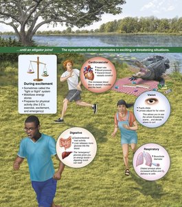
Sympathetic division: Mobilizes the body for activity ("fight or flight" response).
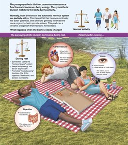
Parasympathetic division: Promotes maintenance functions and conserves energy ("rest and digest" response).
Key Point: Most organs receive dual innervation, allowing precise regulation of physiological functions.
Structural Organization of the ANS
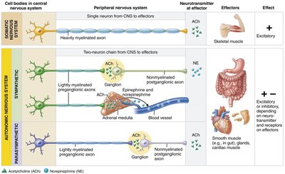
The ANS is part of the motor (efferent) division of the peripheral nervous system. It operates alongside the somatic nervous system but differs in effectors, pathways, and neurotransmitters.
Somatic nervous system: Controls voluntary movements via skeletal muscle.
Autonomic nervous system: Controls involuntary actions via smooth muscle, cardiac muscle, and glands.
Key Anatomical Differences Between ANS Divisions
Origin and Pathways
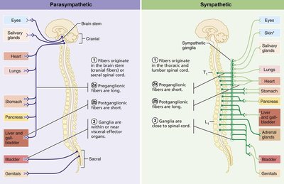
The sympathetic and parasympathetic divisions differ in their anatomical origins, ganglia locations, and fiber lengths.
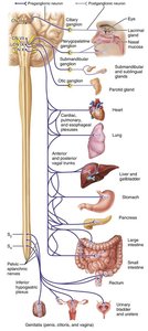
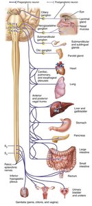
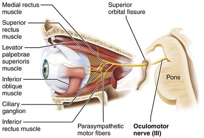
Parasympathetic (craniosacral) division: Originates from brainstem (cranial nerves III, VII, IX, X) and sacral spinal cord (S2–S4).
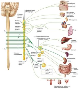
Sympathetic (thoracolumbar) division: Originates from thoracic and upper lumbar spinal cord segments (T1–L2).
Location of Ganglia
Parasympathetic ganglia: Located near or within target organs (terminal or intramural ganglia).
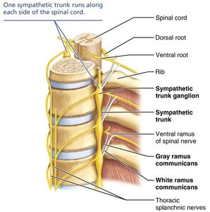
Sympathetic ganglia: Located in sympathetic trunk (paravertebral) ganglia alongside the vertebral column or in collateral (prevertebral) ganglia anterior to the vertebral column.
Fiber Lengths and Branching
Parasympathetic: Long preganglionic, short postganglionic fibers; minimal branching.
Sympathetic: Short preganglionic, long postganglionic fibers; extensive branching.
Functional Roles of the ANS Divisions
Parasympathetic Division: "Rest and Digest"
The parasympathetic division dominates during restful states, promoting digestion, energy storage, and maintenance functions.
Decreases heart rate
Stimulates digestive activity
Promotes urination and defecation
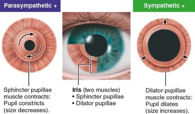
Constriction of pupils
Sympathetic Division: "Fight or Flight"
The sympathetic division prepares the body for emergencies, increasing alertness and metabolic activity.
Increases heart rate and blood pressure
Bronchodilation (widens airways)
Inhibits digestive and urinary functions
Dilates pupils
Stimulates sweat glands and arrector pili muscles
Mobilizes energy stores (glucose, fats)
Dual Innervation and Antagonistic Interactions
Most organs receive input from both divisions, which typically have opposing effects. This antagonism allows for fine-tuned control of organ function.
Example: Sympathetic stimulation increases heart rate, while parasympathetic stimulation decreases it.
Neurotransmitters and Receptors in the ANS
Cholinergic and Adrenergic Transmission
ANS neurons use different neurotransmitters and receptors to communicate with effectors.
Cholinergic fibers: Release acetylcholine (ACh); found in all preganglionic neurons and parasympathetic postganglionic neurons.
Adrenergic fibers: Release norepinephrine (NE); found in most sympathetic postganglionic neurons.
Receptor Types
Nicotinic receptors: Found on all postganglionic neurons (both divisions); always excitatory.
Muscarinic receptors: Found on all parasympathetic target organs; can be excitatory or inhibitory depending on subtype (M1, M2, M3).
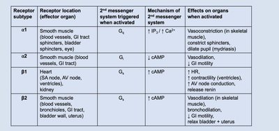
Adrenergic receptors: Found on sympathetic target organs; divided into alpha (α1, α2) and beta (β1, β2) subtypes with varying effects.
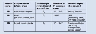
Summary Table: Main Receptor Subtypes and Effects
Receptor subtype | Location | Main Effect |
|---|---|---|
M2 | Heart | Decreases heart rate |
M3 | Smooth muscle, glands | Bronchoconstriction, increased GI motility |
α1 | Blood vessels, GI sphincters | Vasoconstriction, sphincter contraction |
β1 | Heart | Increases heart rate and contractility |
β2 | Bronchioles, GI tract | Bronchodilation, GI relaxation |
Visceral Reflexes
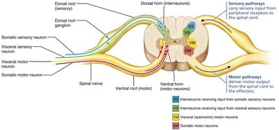
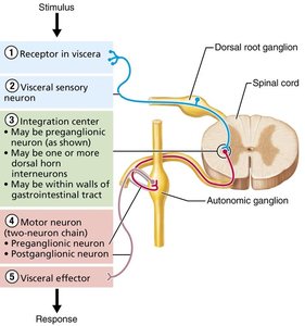
Visceral Reflex Arc
Visceral reflexes control involuntary functions such as heart rate, digestion, and respiratory rate. They involve sensory input from internal organs and motor output via the ANS.
Components: receptor, sensory neuron, integration center, motor neuron (two-neuron chain), effector
Effectors: smooth muscle, cardiac muscle, glands
CNS Control of the ANS
Hierarchy of Control
The ANS is regulated at multiple levels of the central nervous system, including the spinal cord, brainstem, hypothalamus, and cerebral cortex. The hypothalamus is the main integration center for autonomic functions.
Spinal cord: Controls reflexes such as urination and defecation.
Brainstem: Regulates heart rate, blood vessel diameter, gastrointestinal activities, and respiratory rate.
Hypothalamus: Integrates autonomic, endocrine, and somatic responses to maintain homeostasis.
Summary Table: Anatomical and Physiological Differences
Characteristic | Parasympathetic | Sympathetic |
|---|---|---|
Origin | Craniosacral (brainstem, S2–S4) | Thoracolumbar (T1–L2) |
Ganglia location | Near/within target organ | Near spinal cord |
Preganglionic fiber length | Long | Short |
Postganglionic fiber length | Short | Long |
Branching | Minimal | Extensive |
Neurotransmitter (postganglionic) | ACh | NE (except sweat glands: ACh) |
Functional role | Rest and digest | Fight or flight |
Key Examples and Applications
Pupil size: Parasympathetic stimulation constricts pupils; sympathetic stimulation dilates pupils.
Heart rate: Parasympathetic decreases; sympathetic increases.
Bronchioles: Parasympathetic constricts; sympathetic dilates.
Digestive activity: Parasympathetic stimulates; sympathetic inhibits.
Additional info: The ANS is essential for maintaining homeostasis and responding to internal and external stressors. Drugs that mimic or block ANS neurotransmitters are widely used in medicine to treat cardiovascular, respiratory, and gastrointestinal disorders.
