 Back
BackThe Axial and Appendicular Skeleton: Key Bones and Landmarks
Study Guide - Smart Notes
 Tailored notes based on your materials, expanded with key definitions, examples, and context.
The Axial Skeleton
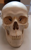
Overview of the Skull
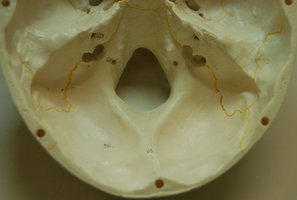
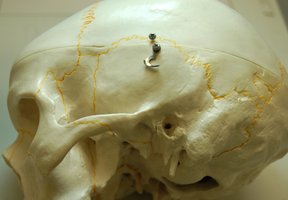
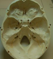
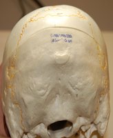
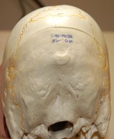
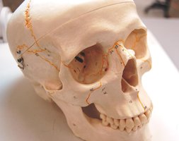
The skull is a complex structure composed of cranial and facial bones that protect the brain and form the structure of the face. It contains numerous foramina and processes that serve as passageways for nerves and blood vessels, as well as attachment points for muscles.
Cranial Bones: Include the frontal, parietal, temporal, occipital, sphenoid, and ethmoid bones.
Facial Bones: Include the maxilla, zygomatic, nasal, lacrimal, vomer, palatine, inferior nasal conchae, and mandible.
Key Landmarks: Supraorbital foramen, infraorbital foramen, mental foramen, optic canal, superior and inferior orbital fissures, and alveolar margins.
Sutures: Coronal, sagittal, lambdoid, and squamous sutures connect the cranial bones.
The Mandible
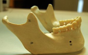
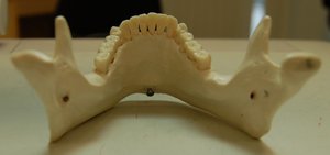
The mandible is the lower jawbone and the only movable bone of the skull. It supports the lower teeth and provides attachment for muscles involved in mastication (chewing).
Key Features: Body, ramus, angle, alveolar margin, mental foramen, mandibular foramen, condyloid process, and coronoid process.
The Vertebral Column
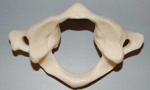
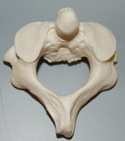
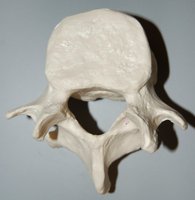
The vertebral column, or spine, is composed of 33 vertebrae divided into cervical, thoracic, lumbar, sacral, and coccygeal regions. It protects the spinal cord and supports the head and body.
Cervical Vertebrae (C1–C7): The atlas (C1) and axis (C2) are specialized for head movement.
Thoracic Vertebrae (T1–T12): Articulate with the ribs.
Lumbar Vertebrae (L1–L5): Largest and support the lower back.
Sacrum and Coccyx: Fused vertebrae forming the base of the spine.
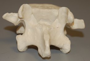
The Thoracic Cage
The thoracic cage consists of the sternum, ribs, and thoracic vertebrae. It protects the heart and lungs and provides attachment for muscles involved in respiration.
Sternum: Composed of the manubrium, body, and xiphoid process.
Ribs: Twelve pairs, classified as true (1–7), false (8–12), and floating (11–12).
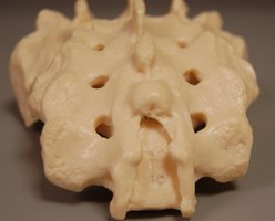
The Appendicular Skeleton
The Pectoral Girdle and Upper Limb
The pectoral girdle consists of the clavicle and scapula, which attach the upper limb to the axial skeleton. The upper limb includes the humerus, radius, ulna, carpals, metacarpals, and phalanges.
Clavicle: Sternal end (medial) and acromial end (lateral).
Scapula: Spine, acromion, coracoid process, glenoid fossa, supraspinous and infraspinous fossae.
Humerus: Head, anatomical neck, greater and lesser tubercles, deltoid tuberosity, capitulum, trochlea, olecranon fossa.
Ulna: Olecranon process, coronoid process, trochlear notch, styloid process.
Radius: Head, radial tuberosity.
Hand: Carpals (scaphoid, lunate, triquetrum, pisiform, trapezium, trapezoid, capitate, hamate), metacarpals, phalanges.
The Pelvic Girdle and Lower Limb
The pelvic girdle is formed by the two hip bones (os coxae), sacrum, and coccyx. The lower limb includes the femur, patella, tibia, fibula, tarsals, metatarsals, and phalanges.
Pelvic Bone: Ilium, ischium, pubis, acetabulum, obturator foramen, pubic symphysis, greater sciatic notch.
Femur: Head, neck, greater and lesser trochanters, linea aspera, medial and lateral condyles.
Tibia: Medial and lateral condyles, tibial tuberosity, anterior margin, medial malleolus.
Fibula: Head, lateral malleolus.
Foot: Tarsals (talus, calcaneus, navicular, cuboid, medial/intermediate/lateral cuneiforms), metatarsals, phalanges.
Summary Table: Major Bones and Landmarks
Region | Key Bones | Major Landmarks |
|---|---|---|
Skull | Frontal, Parietal, Temporal, Occipital, Sphenoid, Ethmoid, Maxilla, Mandible | Foramina, Sutures, Processes |
Vertebral Column | Cervical, Thoracic, Lumbar, Sacrum, Coccyx | Body, Spinous Process, Transverse Process |
Thoracic Cage | Sternum, Ribs | Manubrium, Body, Xiphoid Process |
Pectoral Girdle | Clavicle, Scapula | Acromion, Coracoid Process, Glenoid Fossa |
Upper Limb | Humerus, Radius, Ulna, Carpals, Metacarpals, Phalanges | Tuberosities, Fossae, Processes |
Pelvic Girdle | Ilium, Ischium, Pubis | Acetabulum, Obturator Foramen |
Lower Limb | Femur, Tibia, Fibula, Tarsals, Metatarsals, Phalanges | Trochanters, Condyles, Malleoli |
Additional info: This guide covers the major bones and anatomical landmarks of the axial and appendicular skeleton, as required for foundational understanding in Anatomy & Physiology. Images included are selected for their direct relevance to the described structures and features.
