 Back
BackThe Brain and Cranial Nerves: Structure, Function, and Organization
Study Guide - Smart Notes
 Tailored notes based on your materials, expanded with key definitions, examples, and context.
The Brain: Major Regions and Landmarks
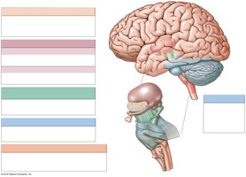
Overview of Brain Organization
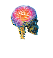
The brain is the central organ of the nervous system, responsible for processing sensory information, regulating bodily functions, and enabling cognition, emotion, and behavior. It is divided into several major regions, each with specialized functions.
Brain Stem: Includes the medulla oblongata, pons, and mesencephalon (midbrain). It controls basic life functions such as heart rate, breathing, and digestion, and serves as a conduit for information between the brain and spinal cord.
Diencephalon: Composed of the epithalamus, thalamus, and hypothalamus. It is involved in sensory processing, hormone production, and autonomic regulation.
Cerebellum: Coordinates voluntary movements and maintains posture and balance.
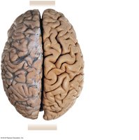
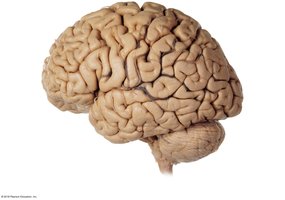
Cerebrum: The largest part of the brain, responsible for higher cognitive functions, voluntary movement, and sensory perception. It is divided into two hemispheres and further into lobes.
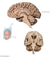
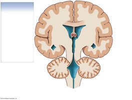
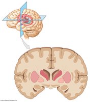
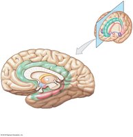
Sectional Anatomy of the Brain
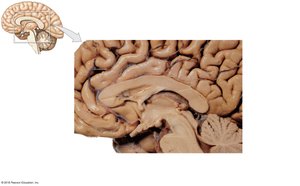
Sagittal and Coronal Sections
Sectional views of the brain provide insight into the internal organization and relationships between different structures. The sagittal section divides the brain into left and right halves, while the coronal section divides it into anterior and posterior parts.
Thalamus: Central relay station for sensory information.
Cerebellum: Located posteriorly, involved in motor coordination.
Medulla Oblongata and Pons: Inferior brainstem structures essential for autonomic functions.
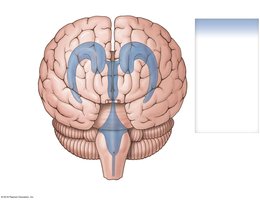
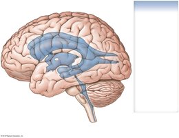
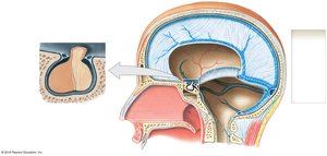
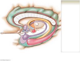
Ventricular System and Cerebrospinal Fluid (CSF)
Ventricles of the Brain
The brain contains four interconnected ventricles filled with cerebrospinal fluid (CSF), which cushions the brain, transports nutrients, and removes waste.
Lateral Ventricles (1 & 2): Located in the cerebral hemispheres; the largest ventricles.
Third Ventricle: Located in the diencephalon.
Fourth Ventricle: Located between the pons and cerebellum; connects to the central canal of the spinal cord.
Cerebral Aqueduct: Connects the third and fourth ventricles.
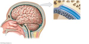
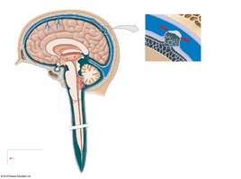
Protection and Support of the Brain
Cranial Meninges
The brain is protected by three connective tissue layers called meninges, as well as the skull and cerebrospinal fluid.
Dura Mater: Outermost, tough layer with periosteal and meningeal components.
Arachnoid Mater: Middle, web-like layer; contains the subarachnoid space filled with CSF.
Pia Mater: Innermost, delicate layer tightly adhering to the brain surface.
Specializations of the Dura Mater
The dura mater forms several folds that help stabilize the brain within the skull:
Falx Cerebri: Separates the two cerebral hemispheres.
Tentorium Cerebelli: Separates the cerebellum from the cerebrum.
Falx Cerebelli: Separates the two cerebellar hemispheres.
Diaphragma Sellae: Covers the pituitary gland in the sella turcica.
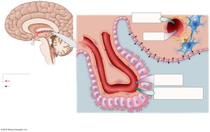
Blood Brain Barrier and CSF Circulation
The blood brain barrier (BBB) is formed by endothelial cells with tight junctions, restricting passage of substances from the blood to the brain. Only lipid-soluble substances can diffuse freely; water-soluble substances require specific transporters. CSF is produced by the choroid plexus and circulates through the ventricles and subarachnoid space, providing nutrients and removing waste.
Brainstem: Medulla Oblongata, Pons, and Mesencephalon
Medulla Oblongata
The medulla oblongata is the lowest part of the brainstem, continuous with the spinal cord. It contains nuclei that regulate autonomic functions (cardiovascular, respiratory, digestive) and relay sensory/motor information. Cranial nerves VIII–XII originate here.
Pons
The pons is a bulge superior to the medulla, containing nuclei for cranial nerves V–VIII, centers for respiratory control, and connections to the cerebellum via cerebellar peduncles.
Mesencephalon (Midbrain)
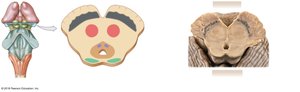
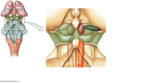
The midbrain contains the corpora quadrigemina (superior and inferior colliculi) for visual and auditory reflexes, as well as the red nucleus and substantia nigra for motor regulation. The cerebral peduncles connect the midbrain to the cerebrum.
Diencephalon
Components and Functions
The diencephalon is composed of the epithalamus (pineal gland), thalamus, and hypothalamus. The pineal gland produces melatonin, the thalamus relays sensory information, and the hypothalamus regulates autonomic and endocrine functions.
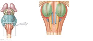
Cerebellum
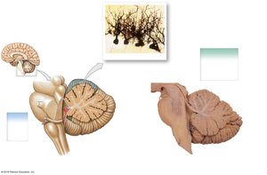
Structure and Function
The cerebellum coordinates voluntary movements and balance. It consists of the cerebellar cortex (gray matter), arbor vitae (white matter), and cerebellar peduncles (fiber tracts connecting to other brain regions). Purkinje cells are large neurons in the cerebellar cortex.
Cerebrum
Major Features
The cerebrum is divided into two hemispheres by the longitudinal fissure and further into lobes: frontal, parietal, occipital, and temporal. It contains gyri (ridges) and sulci (grooves), the corpus callosum (connecting the hemispheres), basal nuclei, and the limbic system.
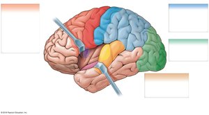
Cerebral Lobes and Functional Areas
Frontal Lobe: Voluntary motor control, planning, and reasoning.
Parietal Lobe: Sensory perception of touch, pressure, pain, temperature, and taste.
Occipital Lobe: Visual processing.
Temporal Lobe: Auditory and olfactory processing.

White Matter Organization
The central white matter of the cerebrum consists of association fibers (within a hemisphere), commissural fibers (between hemispheres), and projection fibers (linking the cerebrum to other brain regions and the spinal cord).
Basal Nuclei
Basal nuclei are clusters of gray matter deep within the cerebral hemispheres. They are involved in the regulation of voluntary motor control, procedural learning, and routine behaviors.
Limbic System
The limbic system is a group of structures involved in emotion, motivation, and memory. It links conscious thought with autonomic functions and facilitates memory storage and retrieval.
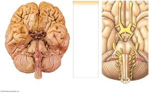
Cranial Nerves
Overview
There are 12 pairs of cranial nerves, each with specific sensory, motor, or mixed functions. They emerge from the brain and innervate structures primarily in the head and neck.
Cranial Nerve | Number | Function |
|---|---|---|
Olfactory | I | Sensory (smell) |
Optic | II | Sensory (vision) |
Oculomotor | III | Motor (eye muscles) |
Trochlear | IV | Motor (superior oblique eye muscle) |
Trigeminal | V | Mixed (sensory: face, motor: mastication) |
Abducens | VI | Motor (lateral rectus eye muscle) |
Facial | VII | Mixed (sensory: taste, motor: facial muscles) |
Vestibulocochlear | VIII | Sensory (hearing, balance) |
Glossopharyngeal | IX | Mixed (sensory: tongue pain, motor: swallowing) |
Vagus | X | Mixed (sensory/motor: organs) |
Accessory | XI | Motor (neck and pharynx muscles) |
Hypoglossal | XII | Motor (tongue muscles) |
Example: The facial nerve (VII) controls facial expressions and conveys taste sensations from the anterior two-thirds of the tongue.
*Additional info: The above content integrates and expands upon the provided lecture slides, adding definitions, examples, and logical academic context for clarity and completeness.*
