 Back
BackThe Cardiovascular System: Blood Vessels and Circulation
Study Guide - Smart Notes
 Tailored notes based on your materials, expanded with key definitions, examples, and context.
The Cardiovascular System: Blood Vessels and Circulation
Overview of Blood Vessels
The cardiovascular system is a closed network of blood vessels that transports blood throughout the body, delivering oxygen and nutrients while removing wastes. Blood vessels are classified into arteries, veins, and capillaries, each with distinct structural and functional characteristics.
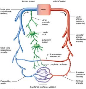
Arteries: Carry blood away from the heart; typically oxygenated except in pulmonary and fetal circulation.
Veins: Return blood to the heart; typically deoxygenated except in pulmonary and fetal circulation.
Capillaries: Microscopic vessels where exchange of gases, nutrients, and wastes occurs between blood and tissues.
Structure of Blood Vessel Walls
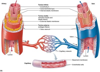
Blood vessel walls (except for capillaries) are composed of three layers, or tunics, each with specialized functions:
Tunica intima: Innermost layer, consisting of endothelium (simple squamous epithelium) that lines the lumen and reduces friction.
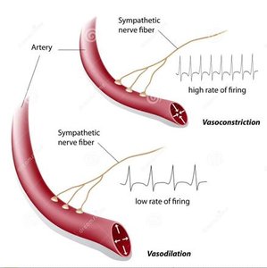
Tunica media: Middle layer, primarily smooth muscle and elastin, responsible for vasoconstriction and vasodilation, thus regulating blood flow and pressure.
Tunica externa (adventitia): Outermost layer, composed of collagen fibers that protect, reinforce, and anchor the vessel to surrounding structures. Large vessels contain vasa vasorum to nourish the external wall.
Types of Blood Vessels
Blood vessels are further classified based on their size, structure, and function:
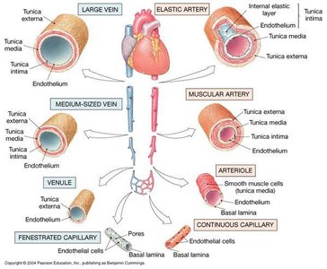
Elastic arteries: Large, thick-walled arteries near the heart; act as pressure reservoirs.
Muscular arteries: Distribute blood to specific organs; more smooth muscle, less elastic tissue.
Arterioles: Smallest arteries; control blood flow into capillary beds via vasoconstriction and vasodilation.
Capillaries: Only endothelium and basal lamina; site of exchange.
Venules: Smallest veins; collect blood from capillaries.
Veins: Larger lumens, thinner walls, contain valves to prevent backflow.
Physiology of Circulation
Key Hemodynamic Terms
Blood flow (F): Volume of blood flowing through a vessel, organ, or the entire circulation per unit time (ml/min). Equivalent to cardiac output for the entire system.
Blood pressure (BP): Force per unit area exerted by blood on vessel walls, measured in mm Hg. Systemic arterial BP is typically measured in the large arteries near the heart.
Resistance (R): Opposition to flow, mainly due to friction between blood and vessel walls. Also called total peripheral resistance (TPR).
Determinants of Resistance
Blood viscosity: Increased viscosity (thicker blood) increases resistance.
Blood vessel length: Longer vessels increase resistance.
Blood vessel diameter: Most important factor; resistance varies inversely with the fourth power of the radius (). Small changes in diameter cause large changes in resistance.
Relationship Between Flow, Pressure, and Resistance
Blood flow is directly proportional to the pressure gradient (ΔP) and inversely proportional to resistance (R):
Thus, increasing pressure increases flow, while increasing resistance decreases flow.
Systemic Blood Pressure
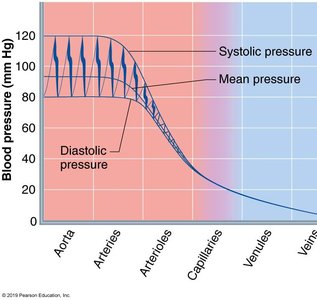
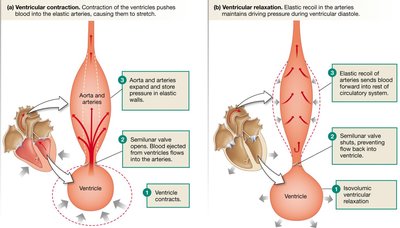
Blood pressure is highest in the aorta and declines throughout the systemic circulation, with the steepest drop in the arterioles. This pressure gradient drives blood flow from arteries to veins.
Arterial Blood Pressure: Systolic, Diastolic, and MAP
Systolic pressure: Peak pressure during ventricular contraction (typically 120 mm Hg).
Diastolic pressure: Lowest pressure during ventricular relaxation (typically 80 mm Hg).
Pulse pressure: Difference between systolic and diastolic pressures.
Mean arterial pressure (MAP): Average pressure that propels blood to tissues. Calculated as:
Example: For BP = 120/80 mm Hg, MAP = 80 + (1/3)(40) = 93 mm Hg.
Clinical Monitoring of Blood Pressure
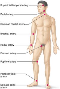
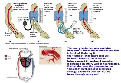
Vital signs include pulse, blood pressure, respiratory rate, and temperature. Pulse is most commonly measured at the radial artery, but other pressure points exist. Blood pressure is measured using a sphygmomanometer and stethoscope, listening for Korotkoff sounds as the cuff is deflated.
Regulation of Blood Pressure
Main Factors Affecting Blood Pressure
Cardiac output (CO)
Peripheral resistance (PR)
Blood volume
BP varies directly with CO, PR, and blood volume.
Short-Term Regulation
Neural controls: The cardiovascular center in the medulla oblongata regulates heart rate and vessel diameter via reflexes from proprioceptors, baroreceptors, and chemoreceptors.
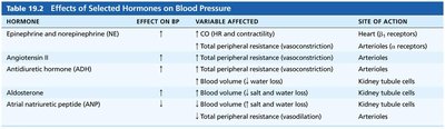
Hormonal controls: Hormones such as epinephrine, norepinephrine, angiotensin II, ADH, aldosterone, and ANP affect blood pressure by altering CO, resistance, or blood volume.
Hormone | Effect on BP | Variable Affected | Site of Action |
|---|---|---|---|
Epinephrine/Norepinephrine | ↑ | CO, TPR | Heart, arterioles |
Angiotensin II | ↑ | TPR (vasoconstriction) | Arterioles |
ADH | ↑ | TPR (vasoconstriction) | Arterioles |
Aldosterone | ↑ | Blood volume | Kidney tubules |
ANP | ↓ | Blood volume, TPR | Kidney tubules, arterioles |
Long-Term Regulation
Long-term regulation is achieved by the kidneys, which control blood volume through direct and indirect mechanisms (renin-angiotensin-aldosterone system).
Capillary Exchange and Fluid Movements
Velocity of Blood Flow
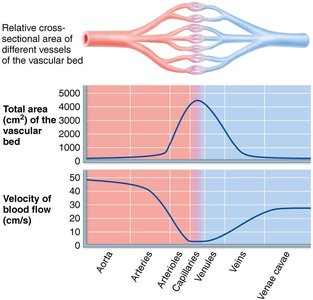
Blood flow velocity is fastest in the aorta and slowest in capillaries, allowing time for exchange. Velocity is inversely related to the total cross-sectional area of the vessels.
Capillary Exchange Mechanisms
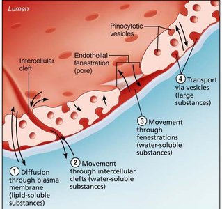
Exchange of gases, nutrients, and wastes occurs via four routes:
Direct diffusion through endothelial membranes (lipid-soluble substances)
Movement through intercellular clefts (water-soluble substances)
Movement through fenestrations (water-soluble substances)
Transport via vesicles or caveolae (large substances like proteins)
Bulk Flow: Filtration and Reabsorption
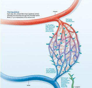
Bulk flow refers to the movement of fluid across capillary walls, driven by hydrostatic and osmotic pressures. This process determines the distribution of fluid between the bloodstream and interstitial space.
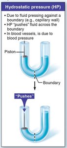
Hydrostatic pressure (HP): The force exerted by fluid pressing against a boundary (e.g., capillary wall). Pushes fluid out of capillaries.
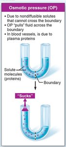
Colloid osmotic pressure (OP): The "sucking" pressure created by nondiffusible plasma proteins, pulling water into capillaries.
Net Filtration Pressure (NFP)
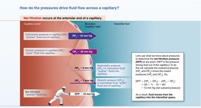
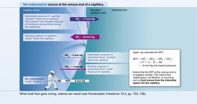
The direction and amount of fluid movement depend on the balance of hydrostatic and osmotic pressures. Net filtration pressure (NFP) is calculated as:
At the arterial end: Net fluid moves out (filtration).
At the venous end: Net fluid moves in (reabsorption).
Excess interstitial fluid is returned to the blood via the lymphatic system.
Venous Blood Pressure and Venous Return
Venous blood pressure is low and changes little during the cardiac cycle. Several mechanisms assist venous return to the heart:
Muscular pump: Skeletal muscle contractions "milk" blood toward the heart.
Respiratory pump: Pressure changes during breathing move blood toward the heart.
Sympathetic venoconstriction: Smooth muscle constriction pushes blood toward the heart.
Clinical Relevance
Monitoring and managing blood pressure is essential for diagnosing and treating cardiovascular diseases such as hypertension and shock.
Understanding blood vessel function is crucial for interpreting drug effects and pharmacokinetics.
Knowledge of circulation helps in patient counseling and recognizing adverse effects of medications.
