 Back
BackThe Cardiovascular System: The Heart – Structure, Function, and Regulation
Study Guide - Smart Notes
 Tailored notes based on your materials, expanded with key definitions, examples, and context.
The Cardiovascular System: The Heart
Overview of Heart Function
The heart is a muscular organ responsible for pumping blood throughout the body via two main circuits: the pulmonary and systemic circuits. It ensures the delivery of oxygen and nutrients to tissues and the removal of carbon dioxide and metabolic wastes.
Heart Structure and Location
Size, Location, and Orientation
Size: Roughly the size of a fist and weighs less than a pound.
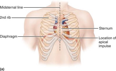
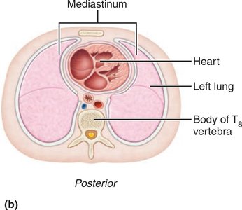
Location: Situated in the mediastinum, between the second rib and fifth intercostal space, on the superior surface of the diaphragm, and about two-thirds to the left of the midsternal line.
Orientation: The base is directed toward the right shoulder, and the apex points toward the left hip. The apical impulse can be palpated between the fifth and sixth ribs, just below the left nipple.
Pericardium and Heart Wall Layers
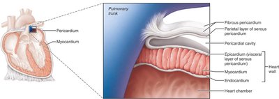
The heart is enclosed in a double-walled sac called the pericardium:
Fibrous pericardium: Superficial layer that protects, anchors, and prevents overfilling.
Serous pericardium: Deep, double-layered membrane (parietal and visceral layers) separated by the pericardial cavity, which contains fluid to reduce friction.
The heart wall consists of three layers:
Epicardium: Visceral layer of serous pericardium.
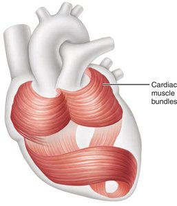
Myocardium: Cardiac muscle layer responsible for contraction; contains a fibrous skeleton for support and electrical insulation.
Endocardium: Endothelial layer lining the heart chambers and valves.
Heart Chambers and Associated Great Vessels
Chambers of the Heart
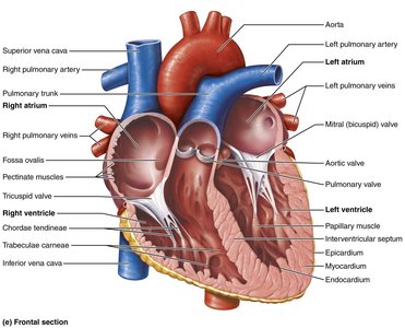
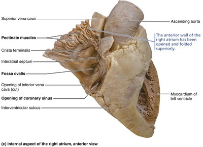
Atria (Right and Left): Superior, thin-walled chambers that receive blood returning to the heart.
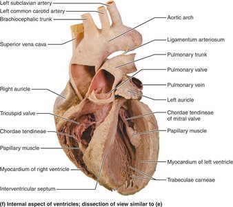
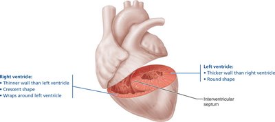
Ventricles (Right and Left): Inferior, thick-walled chambers that pump blood out of the heart.
Septa: Interatrial septum separates atria; interventricular septum separates ventricles.
Major Vessels
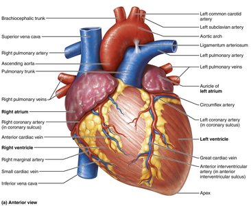
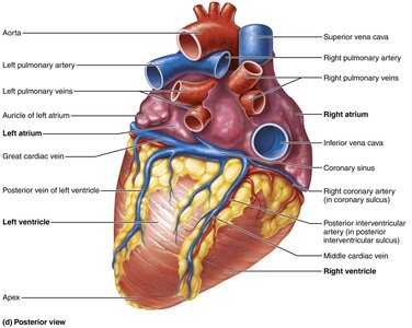
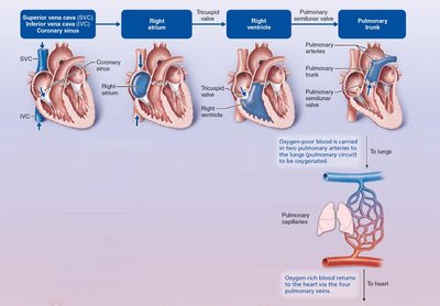
Right atrium: Receives oxygen-poor blood from the superior and inferior vena cava and coronary sinus.
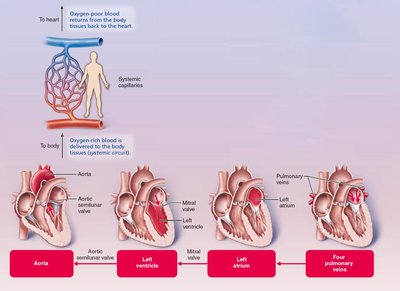
Left atrium: Receives oxygen-rich blood from four pulmonary veins.
Right ventricle: Pumps blood into the pulmonary trunk (to the lungs).
Left ventricle: Pumps blood into the aorta (to the systemic circuit).
Heart Valves and Blood Flow
Heart Valves
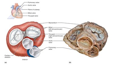
Valves ensure unidirectional blood flow through the heart:
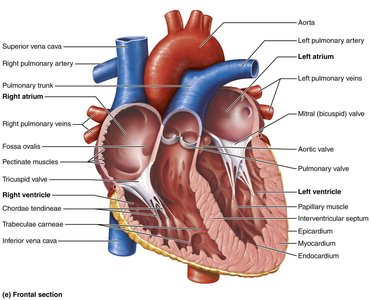
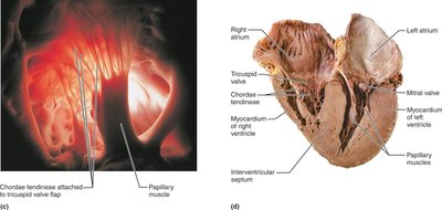
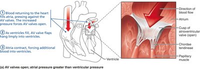
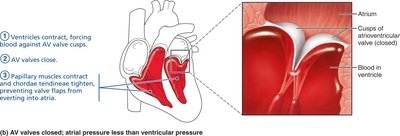
Atrioventricular (AV) valves: Tricuspid (right) and mitral/bicuspid (left) valves between atria and ventricles; prevent backflow into atria during ventricular contraction.
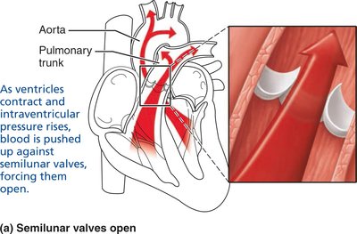
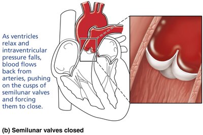
Semilunar (SL) valves: Pulmonary (right) and aortic (left) valves between ventricles and major arteries; prevent backflow into ventricles during relaxation.
Blood Flow Through the Heart
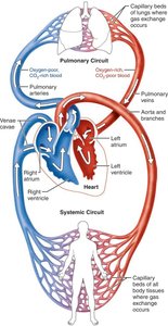
Blood flows from atria to ventricles, then to the lungs or systemic circulation:
Right side: Receives oxygen-poor blood and pumps it to the lungs (pulmonary circuit).
Left side: Receives oxygen-rich blood from the lungs and pumps it to the body (systemic circuit).
Both circuits move equal volumes of blood, but the left ventricle works harder due to higher resistance in the systemic circuit.
Coronary Circulation
Arterial Supply
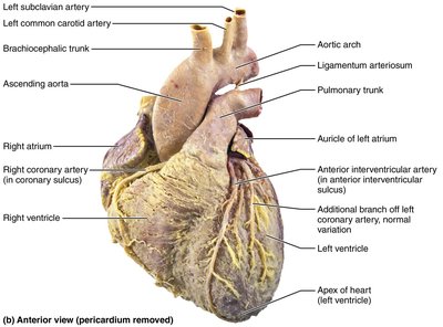
Coronary arteries: Arise from the base of the aorta and supply the heart muscle (myocardium).
Left coronary artery: Branches into the anterior interventricular artery and circumflex artery.
Right coronary artery: Branches into the right marginal artery and posterior interventricular artery.
Venous Drainage
Cardiac veins: Collect deoxygenated blood from the myocardium and drain into the coronary sinus, which empties into the right atrium.
*Additional info: Coronary circulation is essential for delivering oxygen and nutrients to the heart muscle itself. Blockage can lead to myocardial infarction (heart attack).*
