 Back
BackThe Cell: Structure, Function, and Membrane Transport
Study Guide - Smart Notes
 Tailored notes based on your materials, expanded with key definitions, examples, and context.
The Cell: Structure and Function
Introduction to Cells
Cells are the fundamental structural and functional units of all living organisms. In humans, cells vary in size, shape, and function, but all share common features that enable them to maintain life processes.
Cell metabolism: Includes anabolic (building) and catabolic (breaking down) reactions, as well as oxidation-reduction reactions for energy production.
Transport of substances: Cells move compounds internally and externally for various functions.
Communication: Cells use chemical and electrical signals to interact.
Cell reproduction: Cells divide to produce new cells for growth, repair, and reproduction.
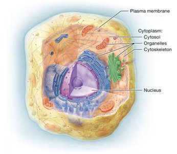
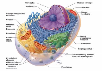
Major Components of the Cell
Plasma membrane: Acts as a selective barrier, providing structural support, communication, regulation of transport, and cellular identification.
Cytoplasm: Contains cytosol (intracellular fluid), organelles, and the cytoskeleton.
Nucleus: Surrounded by a nuclear envelope, contains DNA, and controls cellular activities by directing protein synthesis.
Structure of the Plasma Membrane
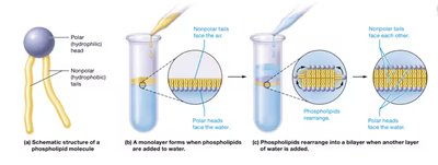
Phospholipid Bilayer
The plasma membrane is primarily composed of a phospholipid bilayer, which forms a barrier between the intracellular and extracellular environments. Phospholipids have both hydrophilic (water-attracting) heads and hydrophobic (water-repelling) tails, allowing the membrane to be selectively permeable.
Polar heads: Face outward toward water-based environments.
Nonpolar tails: Face inward, away from water.
Fluid Mosaic Model
The plasma membrane is described by the fluid mosaic model, which highlights the dynamic arrangement of proteins, lipids, and carbohydrates within the bilayer. This structure allows for flexibility and the presence of various membrane proteins that perform specialized functions.
Integral proteins: Span the membrane and are involved in transport and signaling.
Peripheral proteins: Attached to one side of the membrane, often involved in signaling or structural support.
Carbohydrate chains: Involved in cell recognition and adhesion.

Transport Across the Plasma Membrane
Passive Transport
Passive transport does not require cellular energy (ATP) and relies on concentration gradients to move substances across the membrane.
Simple diffusion: Movement of nonpolar solutes (e.g., gases, lipids) directly through the bilayer.
Facilitated diffusion: Movement of polar or charged solutes (e.g., ions, glucose) via membrane proteins (channels or carriers).
Osmosis: Diffusion of water across a selectively permeable membrane from low to high solute concentration.
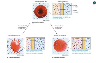
Tonicity
Isotonic solution: No net water movement; cell volume remains constant.
Hypertonic solution: Cell loses water and shrinks (crenation).
Hypotonic solution: Cell gains water and may swell or burst.
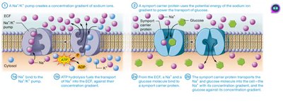
Active Transport
Active transport requires energy (ATP) to move substances against their concentration gradients, often using membrane proteins called pumps.
Primary active transport: Direct use of ATP to transport molecules (e.g., Na+/K+ pump).
Secondary active transport: Uses the energy from the movement of one substance down its gradient to drive the transport of another substance against its gradient.
Vesicular Transport
Large molecules and particles are transported via vesicles in processes requiring ATP:
Endocytosis: Uptake of materials into the cell (includes phagocytosis and pinocytosis).
Exocytosis: Release of materials from the cell.
Cellular Organelles and Their Functions
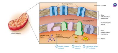
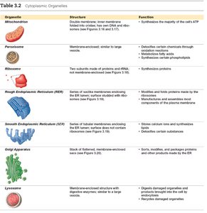
Mitochondria
Mitochondria are the main site of ATP production through oxidative metabolism. They contain their own DNA and ribosomes and are often called the "powerhouses" of the cell.
Ribosomes
Ribosomes are the sites of protein synthesis. They can be free in the cytosol or bound to the rough endoplasmic reticulum (RER).
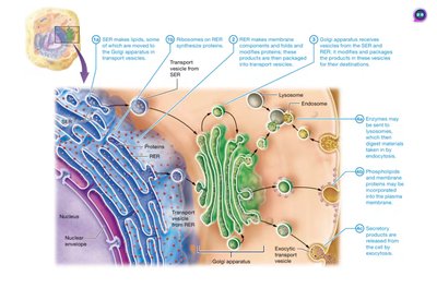
Endoplasmic Reticulum (ER)
Rough ER (RER): Studded with ribosomes; synthesizes and folds proteins, especially those for secretion or membrane insertion.
Smooth ER (SER): Lacks ribosomes; involved in lipid synthesis, detoxification, and calcium storage.
Golgi Apparatus
The Golgi apparatus modifies, sorts, and packages proteins and lipids for delivery to various destinations.

Other Organelles
Organelle | Structure | Function |
|---|---|---|
Mitochondrion | Double membrane, inner membrane folded into cristae | ATP synthesis |
Peroxisome | Membrane-enclosed, similar to large vesicles | Detoxifies chemicals, metabolizes fatty acids |
Ribosome | Two subunits, not membrane-enclosed | Protein synthesis |
Rough ER | Membrane with ribosomes | Protein folding and modification |
Smooth ER | Membrane without ribosomes | Lipid synthesis, detoxification, calcium storage |
Golgi apparatus | Stack of flattened sacs | Sorts, modifies, packages proteins/lipids |
Lysosome | Membrane-enclosed vesicle | Digests damaged organelles and debris |
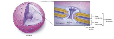
The Nucleus
Structure and Function
The nucleus is surrounded by a double membrane (nuclear envelope) with nuclear pores that regulate the movement of substances between the nucleus and cytoplasm. It contains chromatin (DNA and proteins) and one or more nucleoli, which are sites of ribosome assembly.
Chromatin and Chromosomes
Chromatin: DNA wrapped around histone proteins, loose during interphase.
Chromosomes: Condensed chromatin, visible during cell division.
Protein Synthesis
Protein synthesis involves two main steps:
Transcription: DNA is copied into messenger RNA (mRNA) in the nucleus.
Translation: Ribosomes read mRNA to assemble amino acids into proteins in the cytoplasm.
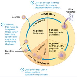
The Cell Cycle
Phases of the Cell Cycle
The cell cycle consists of interphase (G1, S, G2 phases) and the mitotic phase (mitosis and cytokinesis). Interphase is the period of growth and DNA replication, while the mitotic phase is when the cell divides.
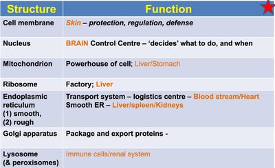
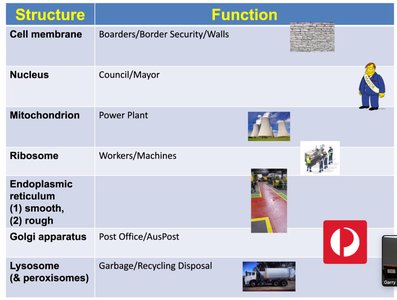
Summary Table: Cell Structures and Functions
Structure | Function |
|---|---|
Cell membrane | Protection, regulation, defense |
Nucleus | Control center, directs activities |
Mitochondrion | ATP production |
Ribosome | Protein synthesis |
Endoplasmic reticulum | Transport system |
Golgi apparatus | Packaging and export |
Lysosome & peroxisome | Digestion and recycling |
Additional info:
Cells communicate via direct passage of molecules or through signaling pathways involving second messengers.
Factors affecting diffusion include concentration gradient, temperature, particle size, diffusion distance, lipid solubility, and membrane permeability.
