 Back
BackThe Central Nervous System: Structure and Function of the Spinal Cord
Study Guide - Smart Notes
 Tailored notes based on your materials, expanded with key definitions, examples, and context.
The Central Nervous System: The Spinal Cord
Introduction
The spinal cord is a critical component of the central nervous system (CNS), serving as the main conduit for information traveling between the brain and the rest of the body. This section explores the anatomical features, protective coverings, internal organization, and functional pathways of the spinal cord, as well as the organization of spinal nerves and nerve plexuses.
Longitudinal Anatomy of the Spinal Cord
Regions and Structure
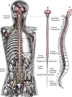
The adult spinal cord is approximately 18 inches long, extending from the foramen magnum of the skull to about the first lumbar vertebra (L1). It is divided into regions that correspond to the vertebral column: cervical, thoracic, lumbar, and sacral. The spinal cord features two enlargements: the cervical enlargement (C3–T2) for nerves to and from the arms, and the lumbar enlargement (T7–T11) for nerves to and from the legs. The spinal cord terminates at the conus medullaris (medullary cone) near L1, and the cauda equina—a bundle of spinal nerves—extends beyond this point. The terminal filum anchors the spinal cord to the coccyx.
31 pairs of spinal nerves emerge from the spinal cord, named according to their region: 8 cervical (C1–C8), 12 thoracic (T1–T12), 5 lumbar (L1–L5), 5 sacral (S1–S5), and 1 coccygeal pair.
The spinal cord does not grow as long as the vertebral column, resulting in the formation of the cauda equina.
The Meninges
Protective Coverings of the CNS
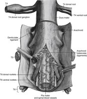
The spinal cord and brain are protected by three connective tissue layers called the meninges:
Dura mater: The tough, outermost layer anchored to the vertebral cavity.
Arachnoid mater: The thin, web-like middle layer, connected to the pia mater by fibrous extensions across the subarachnoid space.
Pia mater: The delicate, innermost layer, rich in blood vessels and in direct contact with the spinal cord.
The subarachnoid space contains cerebrospinal fluid (CSF), which cushions and protects the CNS. CSF can be sampled via a lumbar puncture (spinal tap) for diagnostic purposes, such as detecting meningitis.
Cross-sectional Anatomy of the Spinal Cord
Gray and White Matter Organization
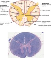
In cross-section, the spinal cord displays a central region of gray matter (shaped like a butterfly or inkblot) surrounded by white matter. The gray matter is divided into horns:
Posterior (dorsal) horn: Receives sensory information from the posterior root.
Anterior (ventral) horn: Contains motor neurons that send signals to skeletal muscles via the anterior root.
Lateral horn: Present in thoracic, upper lumbar, and sacral regions; involved in the sympathetic division of the autonomic nervous system.
The white matter is organized into columns (funiculi):
Posterior columns: Ascending sensory tracts.
Anterior and lateral columns: Both ascending and descending tracts for sensory and motor information.
Other key features include the central canal (containing CSF), gray commissure (connecting sides of gray matter), anterior median fissure, and posterior median sulcus.
Nerve Plexuses
Organization of Peripheral Nerves
Spinal nerves reorganize into nerve plexuses—networks of intersecting nerves—at four main locations along the vertebral column. These plexuses allow axons from different spinal nerves to combine and form peripheral nerves that innervate specific regions of the body.
Cervical plexus (C1–C5): Innervates the neck and diaphragm (phrenic nerve).
Brachial plexus (C5–T1): Innervates the arms (radial, median, ulnar, musculocutaneous, axillary nerves).
Lumbar plexus (L1–L4): Innervates the pelvic region and anterior leg (femoral, obturator nerves).
Sacral plexus (L4–S4): Innervates the posterior leg (sciatic nerve).
Thoracic spinal nerves (T2–T11) do not form plexuses but become intercostal nerves, innervating muscles between the ribs.
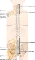
Table: Major Nerve Plexuses and Associated Nerves
Name | Associated Spinal Nerves | Major Associated Peripheral Nerves |
|---|---|---|
Cervical | C1–C5 | Phrenic |
Brachial | C5–T1 | Radial, median, ulnar, musculocutaneous, axillary |
Lumbar | L1–L4 | Femoral, obturator |
Sacral | L4–S4 | Sciatic |
Functional Pathways: Sensory and Motor Signaling
Pathways in the Spinal Cord
Sensory (afferent) signals enter the spinal cord via the posterior (dorsal) root, with cell bodies located in the posterior root ganglion. Motor (efferent) signals exit via the anterior (ventral) root. These pathways are essential for reflexes and voluntary movement, with ascending tracts carrying sensory information to the brain and descending tracts transmitting motor commands to the periphery.
Example: The radial nerve contains both sensory fibers (cutaneous sensation) and motor fibers (muscle movement in the arm).
Clinical Application: Lumbar Puncture and Meningitis
A lumbar puncture is performed in the lower lumbar region to safely withdraw CSF for diagnostic purposes, such as detecting meningitis. Meningitis is an inflammation of the meninges, often caused by infection, and can lead to severe neurological symptoms if untreated.
Key Terms and Definitions
Spinal cord: The main pathway for information connecting the brain and peripheral nervous system.
Meninges: Three protective membranes (dura mater, arachnoid mater, pia mater) surrounding the CNS.
Cauda equina: Bundle of spinal nerves extending beyond the conus medullaris.
Nerve plexus: Network of intersecting nerves formed by the reorganization of spinal nerve axons.
Gray matter: Region of the spinal cord containing neuron cell bodies and synapses.
White matter: Region containing myelinated axons organized into tracts.
Summary Table: Spinal Cord Features
Feature | Description |
|---|---|
Cervical enlargement | Region with increased neural structures for arm innervation (C3–T2) |
Lumbar enlargement | Region with increased neural structures for leg innervation (T7–T11) |
Conus medullaris | Tapered end of the spinal cord (near L1) |
Cauda equina | Bundle of spinal nerves extending beyond the spinal cord |
Central canal | CSF-filled channel running the length of the spinal cord |
Additional info: The spinal cord is essential for reflexes, voluntary movement, and autonomic functions. Damage to the spinal cord can result in loss of sensation, paralysis, or autonomic dysfunction depending on the level and extent of injury.
