 Back
BackThe Digestive System: Structure, Function, and Regulation
Study Guide - Smart Notes
 Tailored notes based on your materials, expanded with key definitions, examples, and context.
The Digestive System
Introduction to the Digestive System
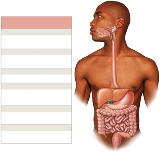
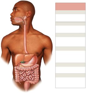
The digestive system is responsible for acquiring nutrients from the environment, synthesizing essential compounds (anabolism), and fueling cellular function through the breakdown of molecules (catabolism). It consists of the digestive tract (gastrointestinal or GI tract) and accessory organs such as teeth, tongue, and various glandular organs.
Digestive tract (alimentary canal): A muscular tube extending from the oral cavity to the anus.
Accessory organs: Teeth, tongue, salivary glands, liver, gallbladder, and pancreas.
Functions of the Digestive System
Ingestion: Entry of food into the oral cavity.
Mechanical digestion and propulsion: Crushing, shearing, and movement of food along the tract.
Chemical digestion: Breakdown of food into small organic fragments for absorption.
Secretion: Release of water, acids, enzymes, buffers, and salts.
Absorption: Movement of nutrients, electrolytes, vitamins, minerals, and water into the body.
Defecation: Elimination of indigestible wastes as feces.
Anatomical Organization and Histology
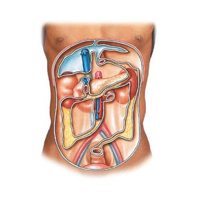
Peritoneal Cavity and Mesenteries
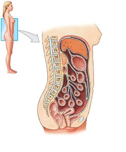
The peritoneal cavity houses most digestive organs and is lined by the peritoneum, a serous membrane with two layers:
Parietal peritoneum: Lines the inner surfaces of the body wall.
Visceral peritoneum: Covers the organs within the cavity.
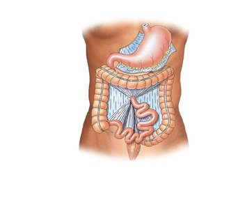
Mesenteries are double layers of peritoneum that suspend and stabilize the digestive tract, provide routes for blood vessels, nerves, and lymphatics, and prevent entanglement of intestines.
Histological Organization of the Digestive Tract
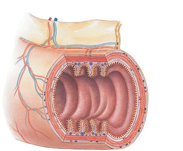
The digestive tract wall consists of four major layers:
Mucosa: Inner lining, a mucous membrane with epithelium, lamina propria, and muscularis mucosae.
Submucosa: Dense irregular connective tissue, may contain exocrine glands.
Muscular layer: Smooth muscle (inner circular and outer longitudinal layers) for mechanical digestion and movement.
Serosa: Outermost serous membrane covering the muscular layer.
Motility and Regulation
Movements in the Digestive System
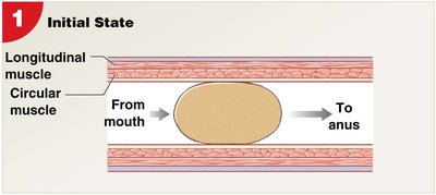
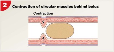
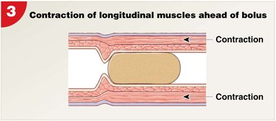
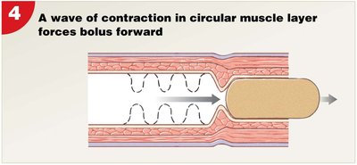
Motility is accomplished by visceral smooth muscle tissue, coordinated by pacesetter cells. Two main types of movement are:
Peristalsis: Waves of muscular contractions that move a bolus along the tract.
Segmentation: Cycles of contraction that churn and fragment the bolus, mixing contents with secretions.
Regulation of Digestive Functions
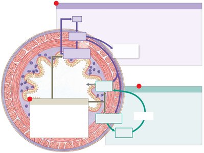
Local factors: pH, volume, and chemical composition in the lumen.
Neural mechanisms: Short (myenteric) and long reflexes coordinate motility and secretion.
Hormonal mechanisms: Peptide hormones produced by enteroendocrine cells regulate digestion.
The Oral Cavity
Anatomy and Functions
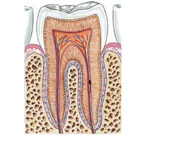
The oral cavity is lined by epithelial tissue, with the hard and soft palates forming the roof and the tongue forming the floor. It contains teeth (incisors, canines, premolars, molars) for mechanical digestion (mastication).
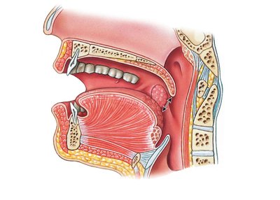
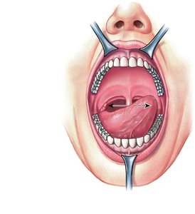
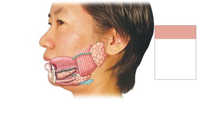
Salivary glands: Produce 1.0–1.5 liters of saliva daily, containing water, electrolytes, buffers, glycoproteins, antibodies, enzymes, and wastes.
Functions of saliva: Cleanses oral surfaces, moistens food, maintains pH, controls bacteria, dissolves chemicals for taste, and initiates carbohydrate digestion with salivary amylase.
The Pharynx and Esophagus
Pharynx
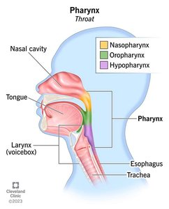
The pharynx is a common passageway for food, liquid, and air, divided into nasopharynx, oropharynx, and laryngopharynx. Food passes through the pharynx to the esophagus during swallowing (deglutition).
Esophagus
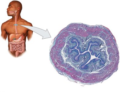
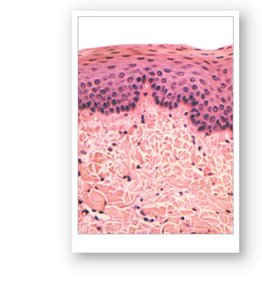
The esophagus is a hollow muscular tube (about 25 cm long) that conveys food and liquids to the stomach. Its resting muscle tone prevents air entry and backflow from the stomach.
Deglutition (Swallowing)
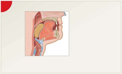
Swallowing involves voluntary and involuntary phases, directing the bolus from the oral cavity to the esophagus and then to the stomach via peristalsis. The gastroesophageal sphincter opens to allow entry into the stomach.
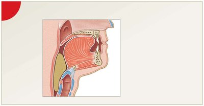
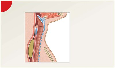
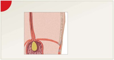
The Stomach
Structure and Function
The stomach temporarily stores ingested food, performs mechanical digestion via muscular contractions, and chemically digests food with acid and enzymes. The bolus becomes chyme, a mixture of partially digested food and gastric secretions.
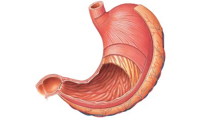
Muscular layer: Includes an additional oblique layer for enhanced mixing and churning.
Mucosa: Simple columnar epithelium with rugae (folds for expansion) and gastric pits leading to gastric glands.
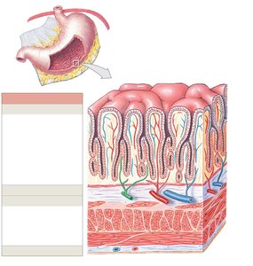
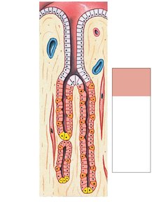
Gastric Gland Cells
Parietal cells: Secrete intrinsic factor (for vitamin B12 absorption) and hydrochloric acid (HCl).
Chief cells: Secrete pepsinogen, activated to pepsin by HCl for protein digestion.
Pyloric glands: Produce mucus; contain enteroendocrine cells (G cells secrete gastrin, D cells secrete somatostatin).
Regulation of Gastric Activity
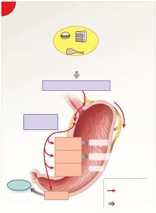
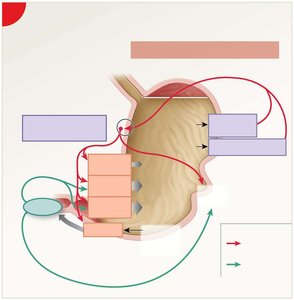
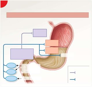
Gastric activity is regulated by the CNS, short reflexes of the enteric nervous system, and digestive hormones. There are three phases:
Cephalic phase: Initiated by sight, smell, taste, or thought of food.
Gastric phase: Triggered by food entry, distension, and chemical changes.
Intestinal phase: Begins as chyme enters the duodenum, regulating gastric emptying.
Accessory Digestive Organs
Pancreas
The pancreas lies posterior to the stomach and has both endocrine (insulin, glucagon) and exocrine (alkaline pancreatic juice with digestive enzymes) functions. Pancreatic juice is delivered to the duodenum via the pancreatic duct.
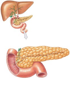
Pancreatic enzymes: Alpha-amylase (starch digestion), lipase (lipid digestion), nucleases (nucleic acid digestion), and proteolytic enzymes (protein digestion).
Liver
The liver is the largest visceral organ, divided into left and right lobes. It performs metabolic and synthetic functions, including nutrient regulation, detoxification, and bile production.
Hepatic portal system: Blood from the digestive tract passes through the liver for nutrient processing and detoxification.
Bile duct system: Bile is secreted by hepatocytes, collected by hepatic ducts, and delivered to the duodenum or stored in the gallbladder.
Gallbladder
The gallbladder stores and concentrates bile, releasing it into the duodenum when stimulated by cholecystokinin (CCK).
The Small Intestine
Structure and Function
The small intestine is a long, muscular tube where chemical digestion is completed and 90% of nutrient absorption occurs. It consists of three segments: duodenum, jejunum, and ileum.
Duodenum: Receives chyme and digestive secretions.
Jejunum: Site of most chemical digestion and absorption.
Ileum: Ends at the ileocecal valve, controlling flow into the large intestine.
Histology of the Small Intestine
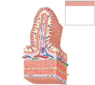
Circular folds: Permanent transverse folds that increase surface area.
Intestinal villi: Fingerlike projections covered by simple columnar epithelium with microvilli (brush border).
Lacteal: Lymphatic vessel in each villus for fat absorption.
Physiology and Regulation
Intestinal juice: Moistens chyme, buffers acids, and keeps enzymes in solution.
Motility: Weak peristaltic contractions move chyme slowly; segmentation mixes contents.
Hormones: Gastrin, secretin, GIP, CCK, VIP, and enterocrinin regulate digestion and absorption.
The Large Intestine
Structure and Function
The large intestine absorbs water, compacts indigestible materials into feces, and stores fecal material prior to defecation. It consists of the cecum, colon (ascending, transverse, descending, sigmoid), and rectum.
Histology: Lacks villi, contains intestinal glands that secrete mucus, and has large lymphoid nodules.
Functions: Absorption of water, vitamins, and bile salts; compaction and storage of feces; houses microbiome that produces vitamins K, biotin, and B5.
Chemical Digestion and Absorption
Digestive Enzymes and Processes
Carbohydrases: Break down carbohydrates.
Proteases: Break down proteins.
Lipases: Break down lipids.
Nucleases: Break down nucleic acids.
Absorption of Nutrients
Carbohydrates: Digested by amylases and brush border enzymes; absorbed as monosaccharides.
Lipids: Digested by lipases; absorbed as fatty acids and monoglycerides, reassembled into chylomicrons, and transported via lacteals.
Proteins: Digested by pepsin, pancreatic proteases, and brush border enzymes; absorbed as amino acids.
Nucleic acids: Digested into nucleotides and absorbed as sugars, phosphates, and nitrogenous bases.
Water: Absorbed passively by osmosis.
Ions: Absorbed by diffusion, carrier-mediated transport, or active transport (e.g., sodium, calcium, potassium, magnesium, iron, chloride, phosphate).
Vitamins: Water-soluble (B, C) and fat-soluble (A, D, E, K) vitamins are absorbed in the small intestine.
Integration with Other Systems
The digestive system is closely integrated with the nervous, cardiovascular, endocrine, and lymphatic systems. It acts as an endocrine organ, producing hormones and neurotransmitters that regulate not only digestion but also other physiological processes.
