 Back
BackThe Digestive System: Structure, Function, and Regulation
Study Guide - Smart Notes
 Tailored notes based on your materials, expanded with key definitions, examples, and context.
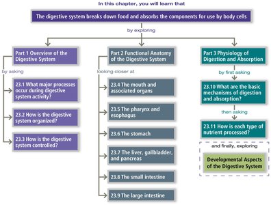
The Digestive System: Overview
Introduction to the Digestive System
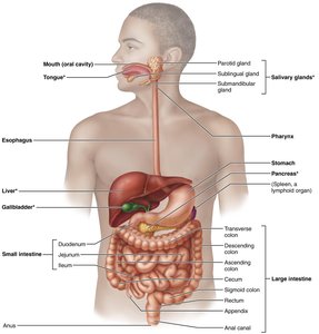
The digestive system is responsible for breaking down food into absorbable components and eliminating indigestible remains. It consists of the alimentary canal (gastrointestinal tract) and accessory digestive organs. The system is organized to maximize nutrient absorption and protect the body from harmful substances.
Alimentary canal: Mouth, pharynx, esophagus, stomach, small intestine, large intestine, rectum, and anus.
Accessory organs: Teeth, tongue, salivary glands, liver, gallbladder, and pancreas.
Major Functions of the Digestive System
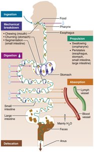
The digestive system performs six essential activities to process food:
Ingestion: Taking food into the mouth.
Mechanical breakdown: Chewing, mixing, and churning food to increase surface area.
Propulsion: Moving food through the GI tract via swallowing and peristalsis.
Chemical digestion: Enzymatic breakdown of food into monomers.
Absorption: Transport of digested nutrients into blood or lymph.
Elimination/Defecation: Removal of indigestible substances as feces.
Protection and Regulation
The GI tract is exposed to the external environment and requires robust protective mechanisms, including digestive enzymes, immunoglobulins, lysozyme, defensins, acidic pH, macrophages, and MALT (mucosa-associated lymphoid tissue). Regulation is achieved through hormonal and neural influences, including the enteric nervous system ("gut brain").
Anatomy of the Digestive System
Gross Anatomy
The digestive tract is a continuous tube with specialized regions for different functions. Accessory organs assist in digestion by producing secretions that enter the GI tract.
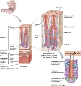
Histology of the Alimentary Canal
The wall of the alimentary canal consists of four basic layers (tunics):
Mucosa: Innermost layer; secretes mucus, digestive enzymes, and hormones; absorbs end products; protects against infection.
Submucosa: Connective tissue with blood vessels, nerves, and lymphatics.
Muscularis externa: Smooth muscle responsible for segmentation and peristalsis.
Serosa: Outermost layer; visceral peritoneum.
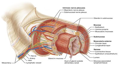
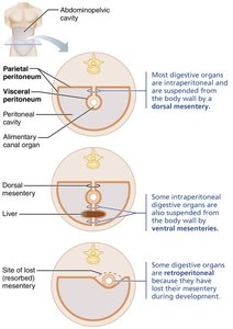
Peritoneum and Mesenteries
The peritoneum is a serous membrane lining the abdominal cavity. Mesenteries are double layers of peritoneum that support and anchor digestive organs, provide pathways for blood vessels, nerves, and lymphatics, and store fat.
Functional Anatomy of the Digestive System
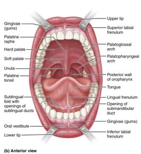
The Mouth and Associated Organs
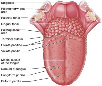
The mouth (oral cavity) is the entry point for food and is lined with stratified squamous epithelium to resist abrasion. It contains the teeth, tongue, and salivary glands, which initiate mechanical and chemical digestion.
Teeth: Mechanically break down food (mastication).
Tongue: Mixes food, forms the bolus, and initiates swallowing.
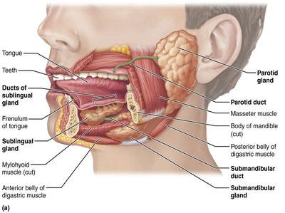
Salivary glands: Produce saliva containing enzymes (e.g., amylase) for starch digestion.
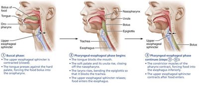
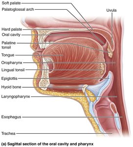
Pharynx and Esophagus
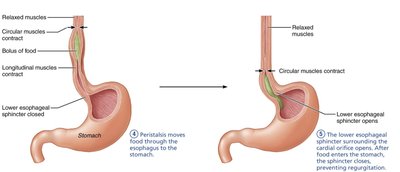
The pharynx and esophagus serve as conduits for food passage from the mouth to the stomach. Swallowing (deglutition) involves coordinated muscle contractions to move the bolus through the pharynx and esophagus via peristalsis.
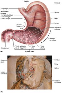
The Stomach
The stomach is a muscular organ where protein digestion begins and food is converted to chyme. It has specialized regions (cardiac, fundus, body, pyloric) and features rugae for expansion. The muscularis externa includes an additional oblique layer for churning food.

Gastric glands: Secrete mucus, hydrochloric acid, pepsinogen, intrinsic factor, and hormones (e.g., gastrin).
Mucosal barrier: Protects the stomach lining from self-digestion.

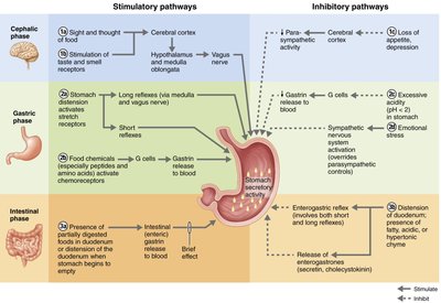
Regulation of Gastric Secretion
Gastric secretion is regulated by neural and hormonal mechanisms in three phases:
Cephalic phase: Triggered by sight, thought, or smell of food.
Gastric phase: Initiated by food entry, stomach distension, and chemical stimuli.
Intestinal phase: Begins as chyme enters the duodenum; includes both excitatory and inhibitory components.
Mechanism of Hydrochloric Acid Secretion
Hydrochloric acid (HCl) secretion by parietal cells is stimulated by acetylcholine, histamine, and gastrin, each acting through different second messenger systems. The combined effect of all three ligands produces maximal acid secretion. HCl is essential for protein digestion and defense against pathogens.
Equation for HCl production:
The Small Intestine
Gross and Microscopic Anatomy
The small intestine is the primary site for digestion and absorption. It is divided into the duodenum, jejunum, and ileum. Structural modifications (plicae circulares, villi, microvilli) greatly increase the surface area for absorption.
Plicae circulares: Circular folds of mucosa and submucosa.
Villi: Fingerlike projections of the mucosa.
Microvilli: Tiny projections on enterocytes forming the brush border.
Specialized cells include absorptive enterocytes, goblet cells, enteroendocrine cells (secreting CCK and secretin), Paneth cells (antibacterial), and stem cells. Peyer's patches (MALT) provide immune protection.
The Liver, Gallbladder, and Pancreas
Liver
The liver is the largest gland in the body, with four lobes and specialized structures such as the falciform ligament and ligamentum teres. Its main functions include processing nutrients, detoxification, storage of vitamins, and production of bile.
Liver lobules: Hexagonal units composed of hepatocytes radiating from a central vein.
Portal triads: Consist of a bile duct, hepatic arteriole, and hepatic portal venule.
Kupffer cells: Macrophages in liver sinusoids.
Bile produced by hepatocytes is essential for fat emulsification and absorption.
Gallbladder
The gallbladder stores and concentrates bile, releasing it into the duodenum via the cystic duct and bile duct in response to cholecystokinin (CCK).
Pancreas
The pancreas has both exocrine (digestive enzyme production) and endocrine (insulin and glucagon secretion) functions. Pancreatic juice contains enzymes for all macromolecules and bicarbonate to neutralize acidic chyme.
Enzymes: Amylase, lipases, nucleases, and proteases (trypsin, chymotrypsin, carboxypeptidase).
Regulation: CCK and secretin stimulate pancreatic secretion in response to chyme entering the duodenum.
The Large Intestine
Structure and Function
The large intestine absorbs water, electrolytes, and vitamins, and compacts indigestible material into feces. It consists of the cecum, appendix, colon (ascending, transverse, descending, sigmoid), rectum, and anal canal. The bacterial flora ferment indigestible carbohydrates and synthesize vitamins B and K.
Chemical Digestion and Absorption
Carbohydrates
Enzymes: Salivary amylase, pancreatic amylase, brush border enzymes.
Absorption: Cotransport with Na+ and facilitated diffusion into capillaries, then transported to the liver via the hepatic portal vein.
Proteins
Enzymes: Pepsin (stomach), trypsin, chymotrypsin, carboxypeptidase (pancreas), brush border peptidases.
Absorption: Similar to carbohydrates, via active transport and facilitated diffusion.
Fats
Emulsification: Bile salts break fat globules into smaller droplets.
Digestion: Pancreatic lipase produces monoglycerides and free fatty acids.
Absorption: Formation of micelles, diffusion into enterocytes, formation of chylomicrons, entry into lacteals, and transport via lymph.
Nucleic Acids
Enzymes: Pancreatic ribonuclease and deoxyribonuclease, brush border nucleosidases and phosphatases.
Absorption: Active transport into enterocytes and transport to the liver.
Electrolytes and Water
Electrolytes: Actively absorbed along the small intestine; sodium is coupled with glucose and amino acid absorption; calcium absorption is regulated by vitamin D and parathyroid hormone.
Water: Absorbed by osmosis, primarily in the small intestine; water uptake is coupled with solute uptake.
Summary Table: Functions of Gastrointestinal Organs
Organ | Major Functions | Comments/Additional Functions |
|---|---|---|
Mouth and accessory organs | Ingestion, mechanical breakdown, propulsion, chemical digestion (starch) | Saliva moistens food, begins starch digestion, cleanses mouth |
Pharynx and esophagus | Propulsion (swallowing, peristalsis) | Move food to stomach; mucus lubricates passage |
Stomach | Mechanical breakdown, chemical digestion (proteins), absorption (few substances) | Secretes intrinsic factor for vitamin B12 absorption |
Small intestine and accessory organs | Chemical digestion (all macromolecules), absorption | Bile and pancreatic juice enter duodenum; brush border enzymes complete digestion |
Large intestine | Absorption of water, electrolytes, vitamins; propulsion; defecation | Bacterial flora ferment fiber, synthesize vitamins; forms feces |
