 Back
BackThe Endocrine System: Structure, Function, and Clinical Relevance
Study Guide - Smart Notes
 Tailored notes based on your materials, expanded with key definitions, examples, and context.
The Endocrine System
Overview of the Endocrine System
The endocrine system is a network of glands that produce and secrete hormones to regulate various bodily functions, including growth, metabolism, and reproduction. Hormones act as chemical messengers, traveling through the bloodstream to target organs and tissues.
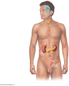
Major endocrine glands: Pineal gland, hypothalamus, pituitary gland, thyroid gland, parathyroid glands, thymus, adrenal glands, pancreas, and gonads (ovaries and testes).
Functions: Regulation of metabolism, growth and development, tissue function, sexual function, reproduction, sleep, and mood.
The Pituitary Gland (Hypophysis)
Anatomy and Structure
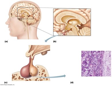
The pituitary gland, often called the "master gland," is located at the base of the brain and is divided into two main lobes: the anterior lobe (adenohypophysis) and the posterior lobe (neurohypophysis). Each lobe releases different hormones that regulate various physiological processes.
Anterior lobe: Produces hormones that regulate growth, metabolism, and reproductive functions.
Posterior lobe: Stores and releases hormones produced by the hypothalamus.
Hormones of the Anterior Pituitary
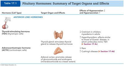
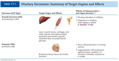
The anterior pituitary secretes several key hormones, each with specific target organs and physiological effects. Disorders can result from either hyposecretion or hypersecretion of these hormones.
Hormone (Cell Type) | Target Organ and Effects | Effects of Hyposecretion and Hypersecretion |
|---|---|---|
Thyroid-stimulating hormone (TSH) | Stimulates thyroid gland to release thyroid hormones | Hypo: Cretinism in children, myxedema in adults Hyper: Hyperthyroidism, Graves' disease |
Adrenocorticotropic hormone (ACTH) | Stimulates adrenal cortex to release glucocorticoids and androgens | Hypo: Rare Hyper: Cushing's disease |
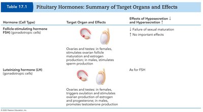
Hormone (Cell Type) | Target Organ and Effects | Effects of Hyposecretion and Hypersecretion |
|---|---|---|
Follicle-stimulating hormone (FSH) | Ovaries and testes: stimulates ovarian follicle maturation and estrogen production in females; stimulates sperm production in males | Hypo: Failure of sexual maturation Hyper: No important effects |
Luteinizing hormone (LH) | Ovaries and testes: triggers ovulation and stimulates ovarian production of estrogen and progesterone in females; promotes testosterone production in males | As for FSH |
Hormone (Cell Type) | Target Organ and Effects | Effects of Hyposecretion and Hypersecretion |
|---|---|---|
Growth hormone (GH) | Liver, muscle, bone, cartilage, and other tissues: stimulates growth, mobilizes fats, increases blood glucose | Hypo: Pituitary dwarfism in children Hyper: Gigantism in children, acromegaly in adults |
Prolactin (PRL) | Breast secretory tissue: promotes lactation | Hypo: Poor milk production in nursing women Hyper: Inappropriate milk production, impotence in males |
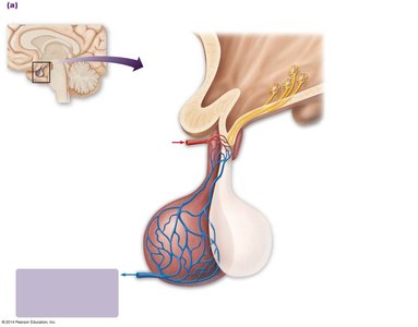
Hypothalamic Control of the Pituitary Gland
The hypothalamus regulates the pituitary gland through two distinct mechanisms: the hypophyseal portal system for the anterior pituitary and the hypothalamo-hypophyseal tract for the posterior pituitary.
Anterior pituitary: Hypothalamic hormones are released into the hypophyseal portal system, stimulating or inhibiting anterior pituitary hormone release.
Posterior pituitary: Hormones (oxytocin and ADH) are synthesized in the hypothalamus and transported down axons to be stored and released from the posterior pituitary.
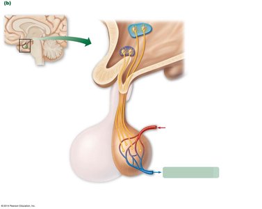
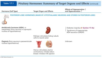
Hormones of the Posterior Pituitary
The posterior pituitary stores and releases two hormones produced by the hypothalamus: antidiuretic hormone (ADH) and oxytocin.
Hormone (Source) | Target Organ and Effects | Effects of Hyposecretion and Hypersecretion |
|---|---|---|
Antidiuretic hormone (ADH) | Kidneys: stimulates kidney tubule cells to reabsorb water | Hypo: Diabetes insipidus Hyper: Syndrome of inappropriate ADH secretion (SIADH) |
Oxytocin | Uterus: stimulates uterine contractions; Breast: initiates milk ejection | Unknown |
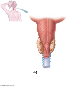
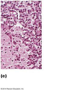
The Thyroid and Parathyroid Glands
Thyroid Gland
The thyroid gland is located in the anterior neck, inferior to the larynx. It produces thyroid hormones (T3 and T4) that regulate metabolism and calcitonin, which lowers blood calcium levels.
Structure: Consists of two lateral lobes connected by an isthmus.
Histology: Composed of follicles filled with colloid, surrounded by follicular cells (secrete thyroid hormone) and parafollicular cells (secrete calcitonin).
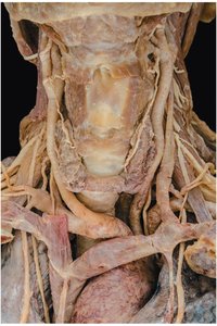
Parathyroid Glands
The parathyroid glands are small glands located on the posterior aspect of the thyroid gland. They secrete parathyroid hormone (PTH), which increases blood calcium levels by stimulating osteoclast activity, increasing intestinal absorption of calcium, and promoting calcium reabsorption in the kidneys.
Histology: Contains chief (parathyroid) cells that secrete PTH and oxyphil cells (function unclear).
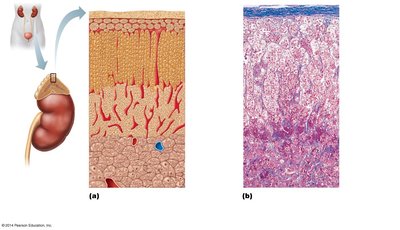
The Adrenal Glands
Structure and Function
The adrenal glands are located atop the kidneys and consist of two regions: the cortex and the medulla. Each region produces different hormones essential for stress response, metabolism, and electrolyte balance.
Adrenal cortex: Produces corticosteroids (mineralocorticoids, glucocorticoids, and androgens).
Adrenal medulla: Produces catecholamines (epinephrine and norepinephrine) for the fight-or-flight response.
Stress Response and the Adrenal Gland
The adrenal gland mediates both short-term and long-term stress responses. The medulla releases catecholamines for immediate effects, while the cortex releases corticosteroids for prolonged stress adaptation.
Short-term stress: Increased heart rate, blood pressure, and metabolic rate; decreased digestive activity.
Long-term stress: Increased blood volume and pressure, protein and fat breakdown, increased blood glucose, suppressed immune system.
Clinical Scenarios and Applications
Case Study: Pituitary Gigantism
A 13-year-old boy presents with rapid growth, headaches, and visual disturbances. Laboratory findings show elevated growth hormone (GH) and a pituitary mass on MRI. This is consistent with pituitary gigantism, caused by excess GH secretion, often due to a pituitary adenoma. Treatment typically involves surgical removal of the tumor, medical therapy to suppress GH, or radiation therapy.
Case Study: Type II Diabetes Mellitus
A 47-year-old woman with obesity is diagnosed with Type II diabetes. The cells not functioning properly are the beta cells of the pancreatic islets, which are responsible for insulin secretion. In Type II diabetes, there is often insulin resistance and eventual beta cell dysfunction. The pancreas is located in the abdomen, posterior to the stomach.
Summary Table: Major Endocrine Glands and Their Hormones
Gland | Main Hormones | Primary Functions |
|---|---|---|
Pituitary (anterior) | GH, TSH, ACTH, FSH, LH, PRL | Growth, metabolism, stress response, reproduction, lactation |
Pituitary (posterior) | ADH, Oxytocin | Water balance, uterine contractions, milk ejection |
Thyroid | T3, T4, Calcitonin | Metabolism, calcium regulation |
Parathyroid | PTH | Calcium regulation |
Adrenal cortex | Corticosteroids (aldosterone, cortisol, androgens) | Electrolyte balance, stress response, metabolism |
Adrenal medulla | Epinephrine, norepinephrine | Fight-or-flight response |
Pancreas | Insulin, glucagon | Blood glucose regulation |
Gonads | Estrogen, progesterone, testosterone | Reproduction, secondary sex characteristics |
Key Concepts and Clinical Relevance
Hormones are chemical messengers that regulate physiological processes.
Feedback mechanisms (mainly negative feedback) maintain hormonal balance.
Disorders can result from hyposecretion (deficiency) or hypersecretion (excess) of hormones.
Understanding endocrine anatomy and physiology is essential for diagnosing and treating related diseases in clinical practice.
