 Back
BackThe Heart and Blood Vessels: Structure, Function, and Circulation
Study Guide - Smart Notes
 Tailored notes based on your materials, expanded with key definitions, examples, and context.
Heart Anatomy and Location
Position and Landmarks
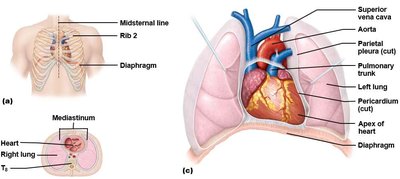
The heart is located obliquely to the midline within the mediastinum, between the lungs. It is anchored by the diaphragm and surrounded by the pericardium. Four anatomical 'corners' help identify its position: superior right, superior left, inferior right, and inferior left. The sternal angle and rib counting are used to locate these landmarks.
Pericardium
The pericardium is a double-walled sac that encloses the heart, providing protection and anchorage. It consists of:
Fibrous pericardium: Anchors the heart to the diaphragm and great vessels.
Serous pericardium: Contains two layers (parietal and visceral/epicardium) with a pericardial cavity filled with fluid to reduce friction.

External and Internal Anatomy of the Heart
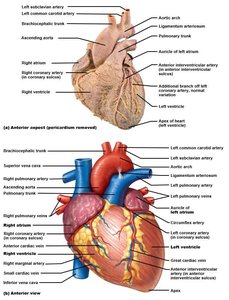
External Features
The heart's surface features include grooves and vessels that help identify its chambers:
Coronary sulcus: Horizontal groove marking the boundary between atria and ventricles.
Anterior and posterior interventricular sulci: Vertical grooves indicating the interventricular septum.
Auricles and great vessels help identify the right and left atria and ventricles.
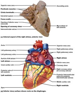
Internal Features
Key internal structures include:
Fossa ovalis: Remnant of the fetal foramen ovale.
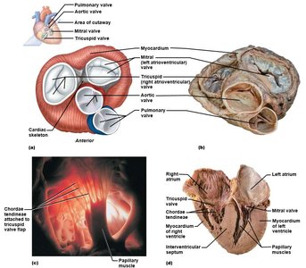
Atrioventricular (AV) valves: Separate atria from ventricles (right: tricuspid, left: bicuspid/mitral).
Semilunar (SL) valves: Separate ventricles from arteries (right: pulmonary, left: aortic).
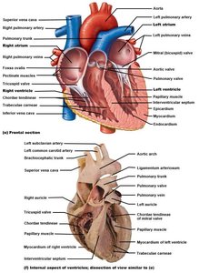
Heart Valves and Their Function
Valve Structure and Function
Heart valves ensure unidirectional blood flow and prevent backflow:
Each valve has 2-3 cusps reinforced by endocardium.
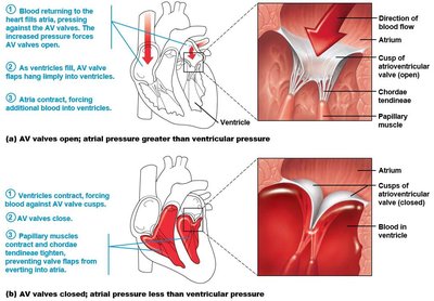
Chordae tendineae connect AV valve cusps to papillary muscles, maintaining closure during ventricular contraction.
Coronary Circulation
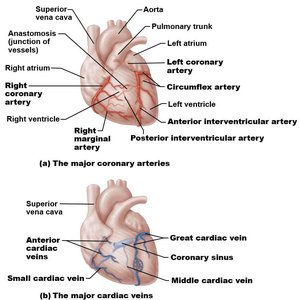
The heart's own blood supply is provided by the coronary arteries and veins:
Left coronary artery (LCA) branches into the anterior interventricular artery (LAD) and circumflex artery (Cx).
Right coronary artery (RCA) branches into the marginal artery and posterior descending artery (PDA).
Venous return is via the coronary sinus, great, middle, and small cardiac veins.
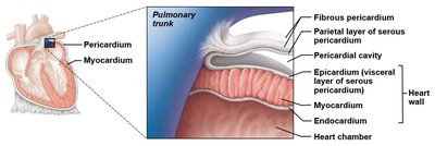
Heart Wall Structure
Layers of the Heart Wall
The heart wall consists of three layers:
Epicardium (visceral pericardium): Outer layer.
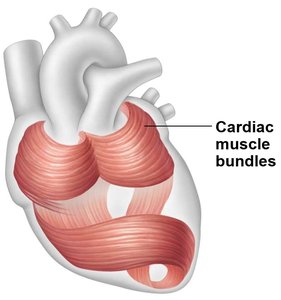
Myocardium: Middle muscular layer, composed of cardiac muscle tissue arranged in spiral and circular bundles. The connective tissue framework is called the cardiac skeleton.
Endocardium: Inner layer, continuous with the endothelium of blood vessels.
Blood Flow Through the Heart
Pathway of Blood
Blood flows through the heart in a specific sequence, ensuring oxygenation and systemic distribution:
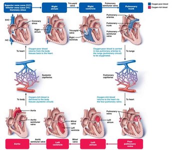
Deoxygenated blood: SVC & IVC → Right atrium → Tricuspid valve → Right ventricle → Pulmonary valve → Pulmonary trunk (to lungs).
Oxygenated blood: Pulmonary veins → Left atrium → Mitral valve → Left ventricle → Aortic valve → Aorta (to body).
Valve Function During Cardiac Cycle
AV valves open during atrial systole/ventricular diastole; close during ventricular systole to prevent backflow.
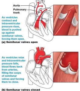
Semilunar valves open during ventricular systole; close during ventricular diastole as blood fills the cusps.
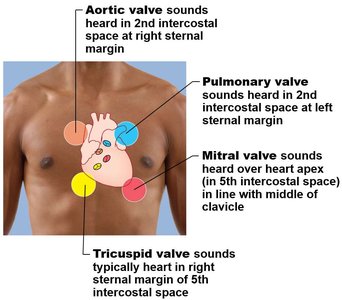
Auscultation Locations
Valve closures create characteristic heart sounds ('lub' for AV valves, 'dupp' for SL valves), best heard at specific chest locations.
Electrical Conduction and Innervation of the Heart
Conduction System
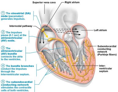
About 1% of myocardial cells are autorhythmic, forming the heart's conduction system:
SA node (pacemaker) → AV node → AV bundle → Bundle branches → Subendocardial conducting network (Purkinje fibers).
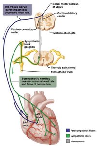
Autonomic Innervation
The heart is innervated by both sympathetic and parasympathetic fibers:
Parasympathetic (vagus nerve): Decreases heart rate.
Sympathetic (cervical and thoracic chain ganglia): Increases heart rate and contraction strength.
Blood Vessels: Structure and Circulation
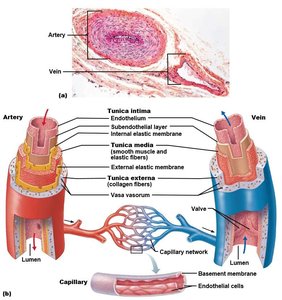
Types and Structure of Blood Vessels
Blood vessels are classified as arteries, veins, or capillaries:
Arteries: Carry blood away from the heart.
Veins: Carry blood toward the heart.
Capillaries: Connect arteries and veins, allowing exchange of gases and nutrients.
Vessel walls have three layers: tunica intima, tunica media, and tunica adventitia.
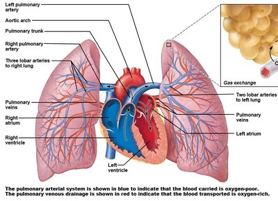
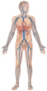
Pulmonary Circulation
Pulmonary circulation exchanges deoxygenated blood for oxygenated blood in the lungs:
Right ventricle → Pulmonary trunk → Pulmonary arteries → Lobar arteries → Arterioles → Capillaries (gas exchange) → Venules → Pulmonary veins → Left atrium.
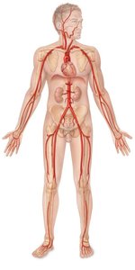
Systemic Circulation
Systemic circulation delivers oxygenated blood to the body and returns deoxygenated blood to the heart:
Left ventricle → Aorta → Elastic arteries → Muscular arteries → Arterioles → Capillaries (exchange) → Venules → Veins → Right atrium.
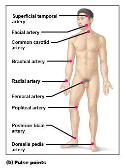
Pulse Points
Pulse points are locations where arteries are superficial and can be palpated to assess heart rate and blood flow.
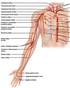
Great Vessels and Vascular Anastomoses
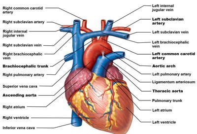
Great Vessels of the Heart
The aorta, superior and inferior vena cavae, and pulmonary arteries and veins are the major vessels entering and leaving the heart. The aortic arch gives rise to the brachiocephalic, left common carotid, and left subclavian arteries.
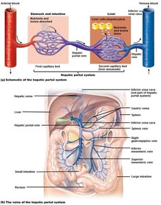
Anastomoses and Portal Systems
Anastomoses are connections between blood vessels that provide alternate routes for blood flow. Portal systems involve two capillary beds in series, such as the hepatic and renal portal systems.
Additional info: The notes above expand on the original content with definitions, context, and examples to ensure a comprehensive, self-contained study guide for college-level anatomy and physiology students.
